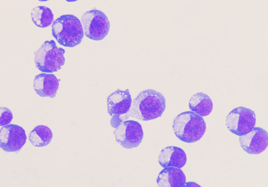

Stem cell transplantation (also called bone marrow transplantation) and gene therapy are among the most powerful treatments for blood diseases such as sickle cell disease, beta-thalassemia, immune deficiencies, and some blood cancers. Replacing or correcting the blood-forming stem cells offers the possibility of long-lasting benefit or a cure.
However, before patients can receive these therapies, they usually need intensive and often toxic chemotherapy or radiation to clear space in the bone marrow for the new stem cells. In a study led by Harvard Medical School researchers at Boston Children’s Hospital and Dana-Farber Cancer Institute published July 8 in Nature, researchers described a new strategy to make stem cell transplants safer by replacing chemotherapy-based treatment with a more targeted approach.
Instead of using toxic agents that damage DNA throughout the body, they used antibodies that recognize specific markers on the blood-forming stem cells in bone marrow. These antibodies can help clear the patient’s existing stem cells in a more selective, less hazardous way.
The work is still in preclinical stages, but limiting or replacing chemotherapy in stem cell transplants could make these types of treatments accessible to new patients.
“By avoiding chemotherapy, we can open up stem cell transplants for diseases that are less severe or for fragile patients normally too sick or too high-risk for transplantation,” said first author Gabriele Casirati, HMS instructor in pediatrics at Boston Children’s.
Molecular camouflage for therapeutic cells
The challenge with using antibodies is that they cannot easily distinguish between the patient’s original stem cells and the infused therapeutic stem cells from the treatment. If the antibody remains in the body, it may also attack the transplanted cells. Senior author Pietro Genovese, HMS assistant professor of pediatrics at Boston Children’s, and his team solved this problem by giving the therapeutic stem cells a form of molecular protection.
Using precise genome-editing tools, they changed a tiny recognition site called an epitope on the surface of the donor stem cells. This small change prevented the antibody from binding to the therapeutic cells without changing their normal function. Essentially, the edited stem cells were given a molecular camouflage: They could hide from the antibody, while the unedited cells remained vulnerable.

The researchers combined the epitope editing with therapeutic edits to increase fetal hemoglobin, a healthy form of hemoglobin that can compensate for the defective adult hemoglobin found in sickle cell disease and beta-thalassemia.
Their results showed that protected stem cells can survive antibody treatment, integrate in the bone marrow, and enrich gradually over time. This creates a new way to not only make room for transplanted cells but also selectively favor the therapeutic cells after transplantation.
Safer, more accessible treatments
This work could help enable chemotherapy-free or chemotherapy-sparing transplantation approaches, reducing the burden of treatment for patients who currently face the risks of DNA damage. And because the antibody can continue to select for protected cells after transplantation, the strategy could help therapeutic stem cells reach the levels needed for clinical benefit.
In previous work published in Nature, the team used the same general principle of epitope editing to protect healthy blood stem cells from powerful cancer immunotherapies, such as CAR T cells or therapeutic antibodies. The therapies attacked leukemia cells and left the blood stem cells alone.
Together, these studies suggest that epitope editing could become a flexible platform. One application could make stem cell transplantation and gene therapy safer, while another could expand the use of cancer immunotherapy by protecting normal blood formation from unintended damage.
“Although this work is still preclinical, it points toward a future in which patients may receive curative stem cell therapies with less toxicity, less reliance on chemotherapy, and greater precision,” said Genovese. “This strategy offers a new framework for safer and more accessible treatments for a wide range of blood diseases.”
Adapted from a Boston Children’s news release.
Authorship, funding, disclosures
Additional authors include Andrea Cosentino, Marta Freschi, Jing Zeng, Adele Mucci, Sébastien Levesque, Nola Neri, Enrico Drago, Viola Carzaniga, Francesco Romano, Varun Katta, Azusa Matsubara, Yichao Li, Mohammed S. Mahmoud, Moisés Chávez-Navarro, Christian Brendel, John P. Manis, Shengdar Tsai, Danilo Pellin, and Daniel Bauer.
This work was supported by the National Institutes of Health (grants R01AI155796, R01HL170629, R01CA286036), the Leukemia Lymphoma Society (grant TRP 6669-24), the Department of Defense (grant HT94252411064), the Alex’s Lemonade Stand Foundation (Research Catalyst 1266935), CURE Childhood Cancer (Translation to CURE 1070087), the Doris Duke Foundation (grant 2022092), St. Jude Children’s Research Hospital Collaborative Research Consortium, the American Society of Transplantation and Cellular Therapy, a DKMS John Hansen Research Grant, the American Society for Gene and Cell Therapy, the PTCTC Jeff Gordon Children’s Foundation, the American Italian Cancer Foundation, the Canadian Institutes of Health Research, an Innovation Fellowship from the Clinical Immunology Society and the Jeffrey Modell Foundation, the Fondo Italiano per la Scienza (FIS00002235), the Italian Leukemia Association sez. Pavia, the Fondazione Enrico ed Enrica Sovena, and the Fondazione Gianni Bonadonna in collaboration with the European School of Oncology.
Casirati, Cosentino, and Genovese are inventors of patent applications related to this work (WO2023159136A2 and WO2024148235A1), which are owned and managed by Boston Children’s and Dana-Farber. The other authors declare no competing interests.