At a glance
Stem cell biologists have developed the first method for growing corticospinal neurons in the lab.
The work advances scientists’ ability to study diseases and injuries that affect those neurons, including amyotrophic lateral sclerosis (ALS) and spinal cord injuries.
The approach also opens new paths for developing regenerative medicine-based treatments.
A team led by Harvard stem cell biologists has discovered a way to grow the type of brain cells that degenerate in patients with amyotrophic lateral sclerosis (ALS) and that are damaged in spinal cord injuries.
Working in lab dishes, researchers engineered a cocktail of molecular signals to coax some progenitor cells — precursors that can differentiate into other cell types — to generate corticospinal neurons, brain cells vital to voluntary motor control.
The work offers the first-ever model for growing corticospinal neurons in the lab.
Although further research is needed to determine whether the approach can be applied to living animal models and human neurons, the current achievement opens new windows for studying and potentially regenerating neurons for the two devastating neurological afflictions.
Findings are published Jan. 27 in the journal eLife.
“The exciting thing about this progenitor population is that it’s already distributed throughout the brain,” said senior author Jeffrey Macklis, the Max and Anne Wien Professor of Stem Cell and Regenerative Biology at Harvard University. “They’re sitting there — resident stem cells.”
A need for regenerative medicine for ALS and spinal cord injuries
In ALS, also known as Lou Gehrig’s disease, corticospinal neurons die for reasons that remain unknown, and eventually patients become paralyzed. The disease afflicts some 30,000 people in the United States.
In spinal cord injuries, the neurons suffer damage when the long axons — which extend up to one meter from the brain to the lower spinal cord — are crushed. About 300,000 Americans currently live with spinal cord injuries.
Many tissues such as skin, bones, and blood can regenerate themselves throughout the lifespan. Most regions in the brain and the spinal cord, however, cannot replace lost or damaged neurons, making neurodegenerative diseases and spinal cord injuries irreversible.

In contrast to embryonic stem cells, which theoretically can give rise to any cell type, progenitor cells are much more advanced on the path to becoming specific cell types with a more limited range of outcomes.
“They’re poised,” said Macklis, who is also Harvard Medical School professor of neurology at Massachusetts General Hospital. “They’ve already gone through a range of developmental steps that we don’t need to orchestrate in a dish or in a brain for regeneration in future experiments.”
Coaxing progenitors into corticospinal neurons
Macklis and colleagues worked with a newly discovered subset of progenitor known as NG2 cells in the cerebral cortex. NG2 cells normally produce oligodendroglia, cells that wrap a fatty substance called myelin around nerve axons that serves like insulation on electrical wires.
Unlike neurons, oligodendrocytes can renew themselves throughout adulthood. For years, biologists have suspected that some unknown subset of these NG2 progenitor cells might retain the dormant capacity to produce neurons. Macklis’s team sought to reawaken this capability.
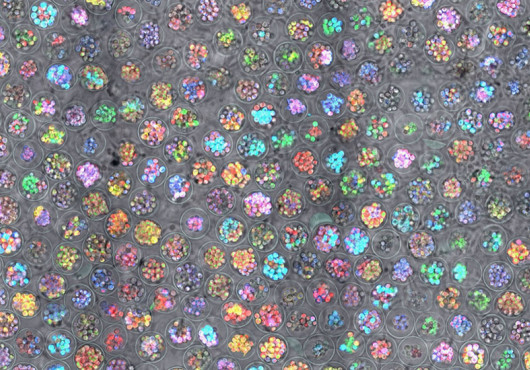
First, researchers purified cultures of cells called SOX6+/NG2+ progenitors. Then they introduced a set of molecular signals, some of which stimulated certain cellular responses and others of which blocked undesired responses, to reproduce how the neurons formed during embryonic development.
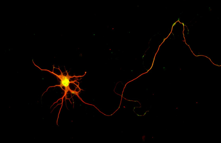
“And lo and behold,” said Macklis, “unlike anything that people have published before, they send out one long axon, and they look like the right kind of neuron.”
Subsequent tests showed that these cells bore all the morphological, molecular, electrophysiological characteristics of normal corticospinal cells and expressed the same genes.
A separate commentary in the journal called the new approach a “perfect recipe” for making these neurons and for reprogramming cells with the goal to repair the brain.
Promise for research and treatment
The work provides the best-yet model for researching the mechanisms of diseases involving corticospinal neurons.
“There are thousands of different types of neurons in the brain,” said co-first author Kadir Ozkan, a former postdoctoral researcher in the Macklis Lab. “Each cell type has a different vulnerability to a given gene mutation. Because of that, we cannot study a disease using just any neuron we are able to generate — we need the right neuron type.”
Further studies should illuminate how to further optimize “the composition of the cocktail [of molecular signals], the dosing, the timing, and all these things, to make it even better,” said co-first author Hari Padmanabhan, also a former postdoctoral researcher in the Macklis Lab.
Potential regenerative therapies that could evolve from the work include transplantation of lab-grown neurons and stimulated neurogenesis in living brains. Macklis said he can foresee such experiments in mice within a few years and, in the more distant future, perhaps humans too.
“If Aladdin came out of the lamp and asked me for my scientific wish,” he said, “my dream experiment would not be to build these neurons in a dish and transplant them. It would to be to bring these types of regulatory controls to bear and activate desired neuron birth and circuit regeneration in situ — right in the brain.”
Adapted from an article in the Harvard Gazette.
Authorship, funding, disclosures
Additional authors include Seth L. Shipman, Eiman Azim, Priyanka Kumar, Cameron Sadegh, and A. Nazli Basak.
This work was supported by the National Institutes of Health (grants NS045523, DP1NS106665, and NS049553), the Emily and Robert Pearlstein Fund for Nervous System Repair, and the Max and Anne Wien Professor of Life Sciences fund.