At a glance
Researchers have answered a longstanding question about how certain non-enveloped viruses punch through the cell membrane to deliver their genetic material.
The findings deepen understanding of virology and could aid the biotech industry’s search for ways to deliver relatively large therapeutic agents into cells to treat or prevent disease.
Work was made possible by an advanced imaging technique known as cryo-electron tomography, or cryo-ET, at HMS.
The future of federally funded research at Harvard Medical School — supported by taxpayers and done in service to humanity — remains uncertain. Learn more.
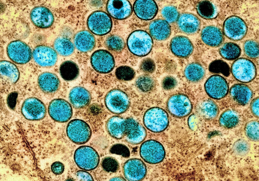
Viruses come in two major flavors — not chocolate and vanilla, quips Harvard Medical School structural biologist Stephen Harrison, but those enveloped in membranes, such as herpesvirus, influenza virus, HIV, and SARS-CoV-2, and those that are not, such as poliovirus, HPV, rotavirus, and adenovirus.
Scientists know more about how enveloped viruses get their genetic material across the cell membrane and into our cells, thereby causing infection, than about how non-enveloped viruses do. Harrison, the Giovanni Armenise-Harvard Professor of Basic Biomedical Sciences at HMS and HMS professor of pediatrics at Boston Children’s Hospital, has spent decades closing this gap.
Thanks to advanced imaging methods, his lab and collaborators have now filled in one of the final pieces of the puzzle for non-enveloped viruses that deliver their genomes packaged within a protein shell.
The findings, supported in part by federal funding and reported Sept. 10 in Science, deepen understanding of virology and could help the biotechnology industry devise better methods to deliver gene therapies and other relatively large therapeutic agents into cells.
“This work is relevant not just for understanding infection by non-enveloped viruses but for addressing the general question of what are the mechanisms for delivering large cargo into cells,” said Harrison, senior author of the study and a Howard Hughes Medical Institute Investigator. “I’ve always thought if we can figure out how evolution did it for one virus, it puts us a step ahead.”
The work was led by first author Marilina de Sautu, research fellow in the Harrison Lab, in close collaboration with Simon Jenni, HMS research scientist in biological chemistry and molecular pharmacology.
How rotavirus punches a hole in the cell compartment
Previous studies from the Harrison Lab and others showed how rotaviruses attach to the cell membrane and then become engulfed and brought into a membrane-bound compartment inside the cell. The virus then must somehow punch a hole in that membrane to get its DNA or RNA into the cell, take over the cell’s molecular machinery, and replicate itself.

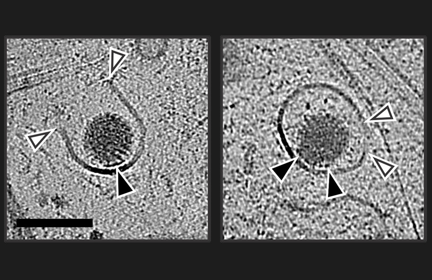
In the new study, which used a strain of rotavirus, de Sautu and colleagues identified the protein that punches the hole in the membrane and revealed how it does so. They showed that virus protein 5, or VP5, makes the membrane permeable to calcium, causing a loss of calcium ions. This in turn makes virus protein 7, or VP7, dissociate from the virus particle and form a pore in the membrane.
To Harrison’s delight, neither the protein nor the mechanism proved to be what he expected.
“All along I had thought it would be VP5 that punched the hole, but it’s VP7,” he said. He noted that work from a former colleague at HMS and Boston Children’s, Philip Dormitzer, who is now a biopharmaceutical executive, and their student, Shane Trask, had hinted that it would be VP7. “This turns out to be hole punching of a particularly satisfying type.”
Made possible by cryo-ET
The work fulfills a career-long goal for Harrison of wanting to see exactly how non-enveloped viruses infect cells, step by step, as a “molecular movie.” The team achieved this using a relatively new technology called cryo-electron tomography, or cryo-ET.
A type of cryo-electron microscopy (cryo-EM), cryo-ET allows scientists to flash-freeze an ultrathin biological sample and take high-resolution snapshots at multiple angles that can be reconstructed into a 3D model. It’s like a CT scan, only instead of the device rotating 180 degrees around a patient to capture an image, in cryo-ET the microscope stays still while the sample tilts.
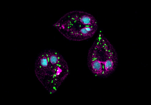
The cryo-ET reconstructions delivered visuals of each step of rotavirus infection, from attaching to the cell membrane and being engulfed to punching the hole and ejecting its RNA. The work was conducted with the help of managing director Richard Walsh and study co-author Conny Leistner at the Harvard Cryo-EM Center for Structural Biology at HMS, for which Harrison serves as faculty director.
Images: Simon Jenni and Marilina de Sautu
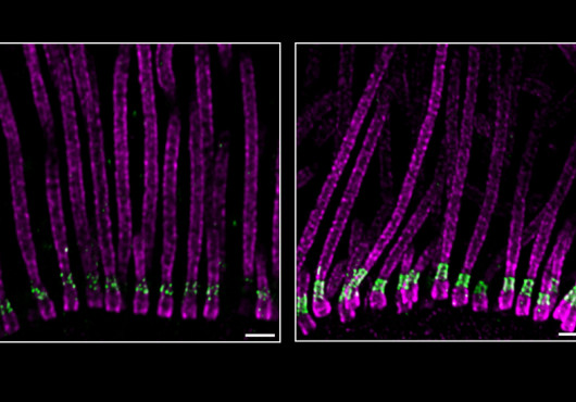
“Cryo-ET allowed us to do something we wouldn’t have been able to do even a few years ago,” said Harrison. “Our paper and [HMS professor] Alan Brown’s work with cilia, published on the same day online in Science, show some of the exciting applications of the technology as HMS applies it to reveal really cool basic biology and address important biomedical problems.”
Live-cell and single-molecule imaging from the lab of co-author Tomas Kirchhausen, professor of cell biology at HMS and HMS professor of pediatrics at Boston Children’s, were critical for motivating and validating the findings.
Future directions
An important set of experiments remains to confirm the specifics of how VP7 works, Harrison said.
Now that the researchers have shown how rotaviruses deliver their genetic material into cells, they plan to study the corresponding mechanisms for certain other non-enveloped viruses. Kirchhausen has already laid the groundwork for imaging the steps for adeno-associated viruses (AAVs), viruses especially relevant to biotech and pharma because they are common choices for delivering gene therapies into cells.
De Sautu plans to investigate other questions relating to viral entry as she completes her postdoctoral research at HMS and starts her own lab.
Both the long-awaited basic-science insight and the potential to assist efforts to treat disease are satisfying, Harrison said.
“Curiosity about how evolution has solved the problem of delivering a large particle into a cell without harming it is my principal motivation,” he said, “but I’m thrilled that it has potential implications from which biotechnologists can take inspiration.”
Authorship, funding, disclosures
This study was funded by the National Institutes of Health (grants R01CA13202, R35GM139386, and R01AI163019) and HHMI.