At a glance
Study reveals how shape changes during the assembly of viral proteins switch on replication in monkeypox virus.
The findings provide a foundation for understanding the molecular machines that drive infection in mpox and aiding the search for therapies.
The future of federally funded research at Harvard Medical School — supported by taxpayers and done in service to humanity — remains uncertain. Learn more.
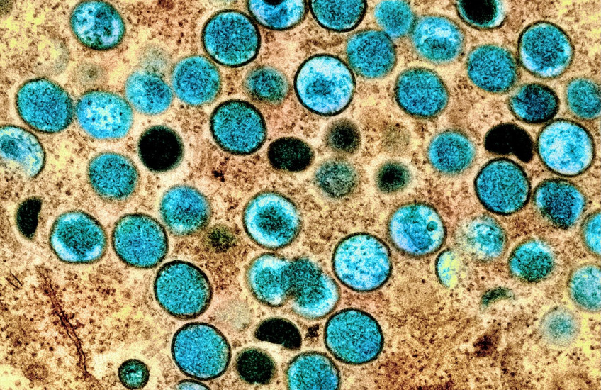
The way a duo of monkeypox virus proteins change shape as they bind together acts like a switch that starts the process of viral replication, a study led by researchers in the Blavatnik Institute at Harvard Medical School reveals.
This kind of detailed structural information promises to help scientists, armed with a new generation of AI-powered virtual screening tools, target their search for ways to inhibit viral replication, treat mpox and related diseases, and prepare for emerging pathogens.
“Learning as much as we can about as many different potential threats as we can is the best way to be ready for the pathogens we will face in the future,” said study co-senior author Jonathan Abraham, professor of microbiology at HMS.
The findings are published Sept. 2 in Nature.
Although vaccines are available to help prevent mpox — the disease caused by the monkeypox virus — and its close relative smallpox, there is currently no antiviral that has been shown to be effective against mpox.
Monkeypox is one in a long and ongoing series of emerging and constantly evolving viral threats to humanity, Abraham said.
The team made its discovery through a combination of two technologies. Cryo-electron microscope imaging showed the structure of the shapeshifting proteins at nearly atomic detail at different moments during assembly and activity, while an exquisitely sensitive tool known as optical tweezers showed how the machinery works in real time to unwind the DNA double helix.
Monkeypox has emerged as a global concern
The monkeypox virus was first discovered in animals in 1958, and the first human case was recorded in 1970. The virus is closely related to smallpox, one of the deadliest viruses humanity has ever faced. Since 2022, an outbreak of mpox has caused more than 100,000 cases in humans and spread to 122 countries, according to the U.S. Centers for Disease Control and Prevention.
Most cases of mpox are relatively mild, but severe cases and complications can cause blindness and even death.
“Mpox is caused by a poxvirus, so we know to be afraid of it,” said Abraham. “There’s a lot that we need to learn to really be prepared for monkeypox virus and other viral threats.”
For example, early in the outbreak that started in 2022, there was hope that the antiviral tecovirimat would be an effective therapy, but it failed to live up to those hopes in a large clinical trial.
Treating patients inspires research
Abraham, a physician-scientist, also works as an infectious disease doctor at Brigham and Women’s Hospital. The challenges he faces in the clinic help inspire his work in the lab. He’s seen firsthand what can happen to people who are sick with viruses for which there is no effective treatment.
While his research is not aimed at finding specific drug targets, Abraham says that broadening and deepening the fundamental understanding of how viruses and other pathogens work is key to accelerating drug discovery.
In this case, he was particularly interested in learning how two key proteins work together to allow monkeypox to replicate itself — the helicase-primase and the polymerase. Each member of the duo performs a distinct task. The helicase-primase unzips the virus’s DNA double helix and attaches a chemical anchor that allows a new strand of DNA to be built, and the polymerase recruits and organizes the building blocks that assemble into a new strand of DNA. Together these pieces are called the replisome.

Previous work by other researchers had defined the structures of these two proteins, but in biochemical tests, the helicase-primase was practically inert. It couldn’t do its job until it worked together with the polymerase.
As a structural biologist who studies how the physical shapes of pathogens, host cell proteins, and other molecules interact in health and disease, Abraham thought that the physical structure of the entire functioning unit might provide useful clues.
“If we want to see how the machinery works, let’s figure out what it looks like, not just in pieces, and not just at rest, but when it’s fully assembled and doing its business,” Abraham said. “Let’s figure out how it moves.”
To see what the functioning replisome looked like, the team used cryo-EM data collected at the Harvard Cryo-EM Center for Structural Biology at HMS. Before the replisome assembles, when the helicase-primase is alone, the primase region of the protein curls up in the center of the helicase, blocking the channel that DNA needs to pass through as it is unwound. But, when the functioning replisome assembles, the polymerase binds to the helicase-primase, pulls the primase region out of the channel, leaving the whole unit open and able to function.
Capturing moments and measuring motion
The cryo-EM images are like a collection of snapshots of the replisome interacting with DNA, offering details of single moments in time.
To confirm that the way they envisioned the process based on those snapshots matched what happens in real time, the researchers used optical tweezers in the lab of co-author Joseph Loparo, professor of biological chemistry and molecular pharmacology at HMS.
The Abraham and Loparo Labs had previously collaborated on a study that used the same techniques to show how a new class of antivirals is able to fight drug-resistant strains of herpes simplex virus.
In this study, the tweezers revealed the moment when the replisome began to unwind the DNA double helix. As hypothesized, when the team added the polymerase to the helicase-primase complex, the fully formed replisome began working.
Challenging research brings potential for great rewards
Having as accurate a model as possible for how these molecular machines operate in real time, with details of how the structures of the component pieces of the replisome change during assembly and activation, gives researchers and increasingly sophisticated artificial intelligence virtual screening tools a much better chance of finding useful therapies and preventive medicines, Abraham said.
Authorship, funding, disclosures
Zishuo Yu is first author of the study. Additional authors include Pradeep Sathyanarayana, Joel M. J. Tan, Side Hu, Xiaoyi Fan, Angela Gao, and Philip J. Kranzusch.
This work was supported by the National Institutes of Health (grants R01 CA272436 and T32 GM145407), a grant from the Cancer Research Institute, a grant by the Broad Institute Center for Integrated Solutions for Infectious Diseases (CISID), and Burroughs Wellcome Fund (BWF) PATH awards. Jonathan Abraham is an investigator of the Howard Hughes Medical Institute.