At a glance
Apicomplexan parasites are a group of pathogens that cause vector-borne diseases, such as babesiosis and malaria.
Researchers have created the most detailed structural map yet of an apicomplexan parasite’s ribosome, the biochemical machinery that makes the cell’s proteins.
The work supports the search for targeted drugs to fight babesiosis, a tick-borne disease affecting humans and livestock.
The study also adds foundational knowledge of this important, understudied group of parasites.
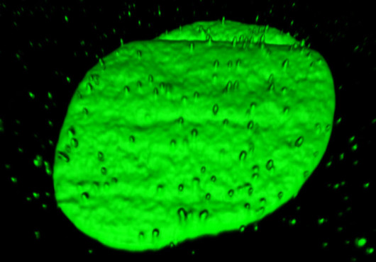
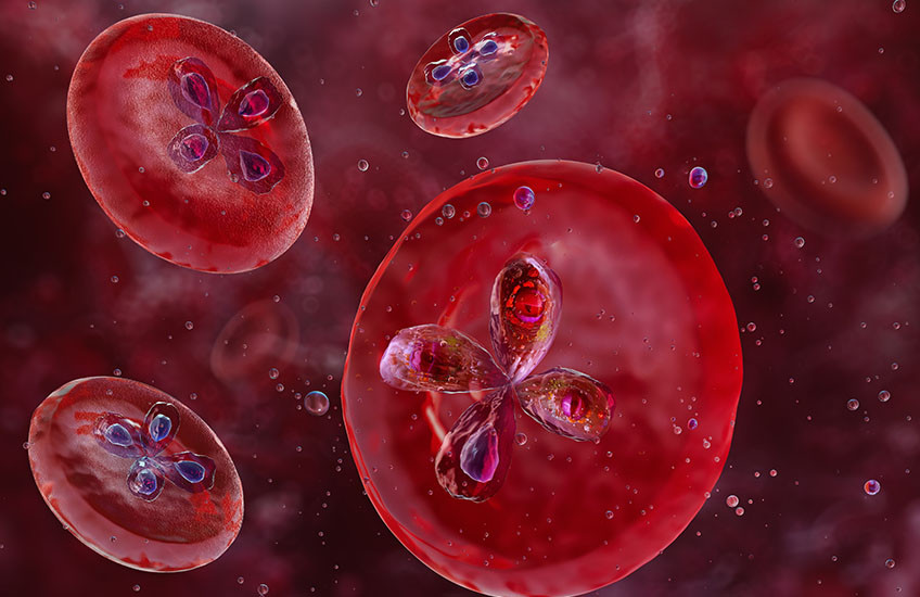
A new study has revealed a three-dimensional, near-atomic-resolution view of the ribosome from Babesia divergens, one of several Babesia species known to infect humans and cause babesiosis. The structural insights from this close-up view could help researchers identify parasite-specific features that may be exploited for future drug development.
The research also deepens the foundations of knowledge for the entire group of protozoa to which Babesia belongs, known as apicomplexan parasites, that are responsible for diseases including malaria and toxoplasmosis. The study published Aug. 6 in Nature Communications.
Very little is known about the fundamental mechanisms that these complex pathogens rely on to cause infection and survive in a diverse range of host species, said Mélissa Léger-Abraham, assistant professor of microbiology in the Blavatnik Institute at Harvard Medical School and HMS assistant professor of pediatrics at Boston Children’s Hospital and senior author of the study.
“Having detailed images of the Babesia ribosome lays an important foundation for deepening our understanding of how the organism lives and gives us step-by-step guidance in the search for new drugs to treat babesiosis,” Léger-Abraham said.
The researchers focused their studies on the ribosome, which is a large molecule composed of ribosomal RNAs (rRNAs) and ribosomal proteins. Ribosomes are cellular biochemical machines that read an organism’s genetic sequence and translate it into proteins that the cell needs to survive. This process, and the ribosome itself, is the target of more than half of the antibiotics currently used against bacterial infections. Therefore, a detailed understanding of the ribosomes of other pathogens, such as parasites, could provide crucial information on how they differ from other organisms like humans or bacteria.
“Once we can see the differences between parasite and human ribosomes, we could design drugs that interrupt the parasite’s life cycle and its ability to infect humans, without interfering with any of the processes that our bodies rely on to keep us healthy,” Léger-Abraham said.
Babesiosis and other tick-borne illnesses are on the rise
Babesia divergens is a tick-borne pathogen prevalent in Europe that causes significant livestock loss and sometimes severe disease — even death — in humans, especially people with compromised immune systems. The disease can spread from one human to another in donated blood and from pregnant mothers to their babies. There are many different species of Babesia that cause this disease in humans. In the United States, cases rose by 9 percent per year from 2015-2022, as the blacklegged ticks that host the closely related Babesia microti have increased their geographic range and the length of their biting season due to warming temperatures.
Léger-Abraham, who has a strong interest in deepening the understanding of neglected tropical diseases, said she was initially interested in imaging ribosomes from Plasmodium, the genus of pathogens that cause malaria, because of its significant global health impact. However, she chose an organism related to Plasmodium, Babesia divergens, because it is easy to cultivate and can produce larger quantities of material required for high-resolution structural studies.
Cryo-EM reveals Babesia’s unique ribosome structure
The researchers used cryo-electron microscopy (cryo-EM) to image a sample of ribosome particles from Babesia divergens. In cryo-EM, many copies of the particles to be analyzed are rapidly frozen in a thin layer of vitreous ice, which is then imaged, providing two-dimensional views of the particles. Researchers then computationally align and combine the resulting two-dimensional images capturing different orientations of the same particles, to reconstruct a three-dimensional model. Data were collected at the Harvard Cryo-EM Center for Structural Biology at HMS.
The structural map Léger-Abraham and colleagues assembled of the Babesia divergens cytosolic ribosomes described the intricate interactions between residues from ribosomal proteins and ribonucleotides from the rRNA. It is the most detailed map of this kind ever generated for an apicomplexan parasite.
Moreover, it is detailed enough to observe chemical modifications of the ribosome that could modulate their function. If you think of RNA as a language, these modifications work like punctuation, accents, and other diacritical marks to subtly modify the meaning of the code.
The team identified 120 of these marks of a type known as rRNA epitranscriptomic marks. These slight chemical modifications to the rRNA can tweak the assembly of the ribosome, which in turn can influence how proteins are made during the translation process. About half of the marks they found sit at positions that had never been reported in any human or other parasite ribosomes. Most are located in the parts of the ribosome that are known to carry out important work, which suggests they are biologically important, and, in some cases, potential drug targets, the authors said.
The researchers did not yet perform follow-up experiments to determine the specific function of the chemical marks that they identified or to see if they have a direct impact on the life cycle of Babesia, but the detailed structure they rendered makes the next steps clear.
“Now that we have a complete list, we can figure out which ones are crucial for infection and/or survival. After that, we can figure out if any of those rRNA modifications are good targets for drug development,” Léger-Abraham said.
Another exciting result was that the researchers were able to detect variations between individual ribosomes. For Babesia, which spends the different stages of its life in very different environments, the differences in the rRNA modifications from one individual to another could offer clues about how the parasite manages its complex life cycle and adapts to survive in diverse hosts, Léger-Abraham said.
Many more puzzles to be solved
In addition to methodically searching for potential drug targets, Léger-Abraham said that another important next step will be imaging the ribosome of the apicoplast, an organelle that performs essential metabolic tasks that Babesia and other apicomplexan parasites need to survive.
This organelle arose through an ancestral endosymbiosis event with a cyanobacterium. Scientists believe an ancestor of the modern protozoans engulfed a blue-green alga, and instead of digesting it, found a way to live together. The apicoplast operates like a semi-autonomous cell within a cell. It has its own DNA genome and its own ribosomes. While Babesia’s main cytoplasmic ribosome is similar to human ribosomes, the apicoplast ribosomes are closer to bacterial ribosomes.
Understanding the unique biology of the apicoplast is another potentially rich resource for discovering antibiotic-like drugs to treat parasitic infections with minimal side effects for humans, Léger-Abraham said.
In addition to pursuing specific investigations to find new therapies, Léger-Abraham said, it’s important to continue curiosity-driven and fundamental research.
“There’s so much data, so much biology, so much information waiting to be revealed that we can use to improve human health,” she said.
Authorship, funding, disclosures
Cristina Gutierrez-Vargas is the first author of the study. Additional authors include Lee S. Izhaki-Tavor, Diana G. Calvopina-Chavez, Caroline D. Keroack, Pablo Copello, and Manoj T. Duraisingh.
This research was supported by the National Institutes of Health’s National Institute of Allergy and Infectious Diseases (grants 5R21AI178196-02, T32AI007061, and 5R01AI67570).