At a glance
Study provides insights into the molecular changes that drive hypertrophic cardiomyopathy (HCM), a thickening and stiffening of heart muscle that can lead to heart failure or sudden cardiac arrest.
The work reveals differences between genetic and non-genetic HCM as well as early and late stages of the disease.
The team identified a gene that may contribute to heart-cell enlargement and found that HCM may affect more cell types than heart muscle cells.
Findings could inform current and future treatments.
The future of federally funded research at Harvard Medical School — supported by taxpayers and done in service to humanity — remains uncertain. Learn more.
An international team led by researchers from Harvard Medical School, Brigham and Women’s Hospital, and the Max Delbrück Center for Molecular Medicine in Germany has created a detailed map of the molecular activity underlying hypertrophic cardiomyopathy (HCM), a common form of heart disease that causes the heart muscle to become thick and stiff and can lead to heart failure or sudden cardiac arrest.
The findings, published Sept. 16 in Science Translational Medicine, provide insights into the changes that drive HCM, which could in turn inform current and future treatments.
“Charting the molecular landscape of hypertrophic cardiomyopathy is essential for advancing our understanding of the disease, improving patient care, and developing new therapeutic approaches,” said co-senior author Christine Seidman, the HMS Thomas W. Smith Professor of Medicine at Brigham and Women’s and professor of genetics at HMS.
“By looking across nearly one million heart cells, we’ve gained important insights into molecular changes that underlie HCM,” added Seidman, who directs the Cardiovascular Genetics Program at the Mass General Brigham Heart and Vascular Institute.
The work reveals differences between genetic and nongenetic HCM as well as early- and late-stage HCM. In fact, the authors said, the findings demonstrate that HCM caused by a genetic defect is distinct from nongenetic HCM, and the molecular changes seen in genetic HCM likely account for more arrhythmias and greater progression to heart failure than nongenetic HCM.
The Seidman Lab — headed by Christine and co-senior author Jonathan Seidman, the Henrietta B. and Frederick H. Bugher Foundation Professor of Genetics in the Blavatnik Institute at HMS — has been a leader in HCM research for decades. Christine Seidman, a practicing cardiologist, brings her patient care experiences back to the lab, where they’re complemented by Jonathan Seidman’s training in genetics. Beginning in 1990, their team made crucial discoveries about the genetic and molecular underpinnings of HCM that paved the way for the first precision treatment for the disease, called mavacamten (Camzyos), approved by the FDA in 2022.
The team behind the new study — co-led by cardiologist Yuri Kim, HMS assistant professor of medicine at Brigham and Women’s, and Eleonora Adami of the Hübner Lab at the Max Delbrück Center — characterized the cellular and molecular signatures of HCM.
The researchers used single-nucleus RNA sequencing to analyze heart tissue from 47 patients spanning early- to end-stage HCM, including people with genetic and nongenetic forms of the disease. They compared these gene expression profiles with data from healthy donor hearts and heart tissue affected by dilated cardiomyopathy, a condition in which the left ventricle balloons, thinning the chamber’s walls.
The data gathered from these nearly one million individual heart cells allowed the creation of the map, which details altered gene activity according to disease stage and genetic status.
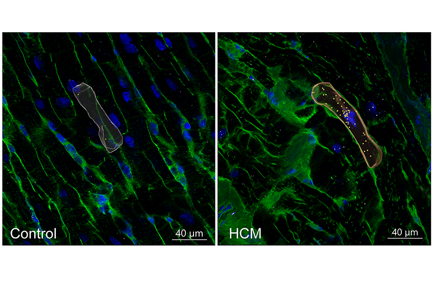
Genetic HCM showed proportionally fewer heart muscle cells than nongenetic HCM and healthy donor tissue. The researchers also identified the gene PRR16 as a potential contributor to cardiomyocyte (heart cell) enlargement that is characteristic of HCM. In addition, genetic HCM samples had elevated expression of genes related to cardiac arrhythmias and fibrosis, features that contribute to disease progression.

The team found distinct features for early-stage HCM compared to late stages of the disease. Fibroblasts — which produce and maintain the extracellular matrix that surrounds cells — expressed less collagen IV, a deficit that the researchers say would likely destabilize the matrix.
In a final step, the researchers showed that an AI model trained on their gene expression data could accurately distinguish early- and late-stage HCM, separate HCM from dilated cardiomyopathy, and correctly identify patients with genetic versus nongenetic HCM. The model was able to make the same distinctions based on gene expression data from fibroblasts alone.
“This was a surprising finding,” said Kim, “because cardiomyopathies are usually thought of as diseases of heart muscle cells only.”
“By mapping gene expression at single-cell resolution across disease stages and genetic subtypes, we’ve built a molecular signature of HCM’s clinical spectrum,” added co-senior author Norbert Hübner at the Max Delbrück Center. “This should provide a foundation for future work on more targeted treatments.”
Adapted from news releases from the Max Delbrück Center and Mass General Brigham.
Authorship, funding, disclosures
Hendrik Milting, Matthias Heinig, Gavin Y. Oudit, and Michela Noseda are also co-senior authors. Additional authors are Sean L. Zheng, Nikolay Shvetsov, Corinna Losert, Henrike Maatz, Syndi Barish, Gabriela Venturini, Natalia López Anguita, Qi Shi, Meraj Neyazi, Martin Beyer, Eric Q. Wei, Amanda Adam, Abhilash Suresh, Daniel Reichart, Eric Lindberg, Kemar J. Brown, Viktoria Strohmenger, David Saul, Anna Gärtner, Michael Lee, Lukas Mach, Jan Lukas Robertus, Joshua M. Gorham, Jan Haas, Laura A. Liebig, Christoph Lippert, Benjamin Meder, Anna Myronova, Giannino Patone, Sam N. Barnett, James S. Ware, Fabio de Robertis, Antonis Pantazis, Jan Gummert, Anissa Viveiros, Huachen Chen, Jorge Ruiz-Orera, Norbert Frey, Barbara A. McDonough, Richard N. Mitchell, Robert F. Padera, Sharlene M. Day, Carolyn Y. Ho, and Neal K. Lakdawala.
This study was supported in part by the Beznos Family Fund; Boehringer Ingelheim Fonds; the British Heart Foundation (BBC/F/21/220106, FS/CRTF/23/24444, SP/19/1/34461, RE/18/4/34215, RE/24/130023); the British Heart Foundation Centre for Research Excellence, Imperial College London; the British Medical Association Foundation Josephine Lansdell Grant; the Deutsches Zentrum für Herz-Kreislauf-Forschung (German Center for Cardiovascular Research) (81Z0600106, 81Z0600105); the Canadian Institutes of Health Research (PJT-451105, PJT-462950); the Chan Zuckerberg Initiative (2019-002431, 2019-202666, 2021-237882); the Erich and Hanna Klessmann Foundation; the German Research Foundation (SFB-1470 Project B03); Imperial College Healthcare NHS Trust Biomedical Research Centre Funding (PA7460); the Medical Research Council UK; the NIHR Imperial Biomedical Research Centre; the National Science Foundation Engineering Research Center on Cellular Metamaterials (EEC-1647837); the Pathfinder Cardiogenomics Programme of the European Innovation Council of the European Union (DCM-NEXT, 101115416); the Rosetrees Trust (PGS23/100028); the Sir Jules Thorn Charitable Trust (21JTA); and the Wellcome Trust (226083/Z/22/Z).
Christine Seidman reports serving as scientific advisor for Maze Therapeutics and Tenaya Therapeutics and on the board of directors for Burroughs Wellcome Fund and Merck. Additional author disclosures can be found in the paper.