At a glance
A new study reveals some of the signals that govern how myelin — a fatty substance that insulates neurons — gets laid down in different amounts in different types of brain cells.
It appears neurons themselves influence the process by communicating with myelin-producing cells called oligodendrocytes.
Findings help illuminate brain development and provide clues that may lead to improved therapies for myelin-related neurological disorders, such as multiple sclerosis.
The future of federally funded research at Harvard Medical School — supported by taxpayers and done in service to humanity — remains uncertain. Learn more.
In the complex circuitry of the brain, a fatty substance known as myelin plays a vital role in insulating neural wiring. It coats billions of axons in the brain and makes up about one-fifth of the organ in humans.
But just how this essential substance is laid down — and how it is customized in diverse types of specialized brain cells — has remained mysterious. By revealing some of the molecular signals that govern this process, a recent study in Developmental Cell by Harvard biologists helps illuminate brain development and provides clues that may lead to therapies for devastating neurological disorders such as multiple sclerosis.
“Knowing how myelination is established is important not only for understanding normal brain development but also for shedding light on neurodevelopmental and neurodegenerative diseases associated with myelin defects,” said first author Nuria Dominguez-Iturza, a postdoctoral researcher in the lab of Paola Arlotta in the Department of Stem Cell and Regenerative Biology at Harvard’s Faculty of Arts and Sciences and Harvard Medical School.
“By understanding how myelination normally occurs, we can better understand what goes wrong in these diseases,” she said.
Different myelination patterns for different neurons
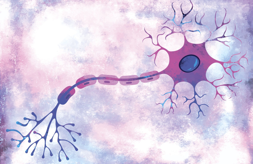
Neurons have long axons that transmit signals to other cells. The longest ones in the human body extend about one meter from the brain to the lower spinal cord. Like electrical wires, they are insulated to facilitate the transmission of signals, in this case by fatty myelin.
In 2014, Arlotta, the Golub Family Professor of Stem Cell and Regenerative Biology at FAS and HMS, and her group published a landmark paper showing that myelination is not a generic, one-size-fits-all process and instead follows varied patterns in different neurons.

For example, some axons are thickly insulated while others have long stretches of unmyelinated tracts. The 2014 study found that myelin distribution was specified by a dialogue between neurons and myelin-producing cells and represented a key feature of neural identity.
What drives the process of this “differential myelination” has remained unknown. The new study sought an answer by probing how neurons interact with myelin-producing cells called oligodendrocytes.
Neurons influence myelin-producing cells
In an investigation that spanned more than four years, Dominguez-Iturza and colleagues examined myelination and myelin-producing oligodendrocytes in mice from early postnatal development to adulthood. Their methods included single-cell RNA sequencing and in utero electroporation, a technique that delivers selected genes to targeted cells in the developing mouse brain.
The team created an interactome — an atlas of the interactions of chemical signals between neurons and oligodendrocytes to see how they play out in different layers of the cortex over time.
The team didn’t find evidence that different types of oligodendrocytes drive differential myelination in the cortex. Instead, they saw that the same types of oligodendrocytes perform differently based on their maturation stage and location in the cortex.
The investigators did find, however, that neurons influence this process through signals to the oligodendrocytes. They showed that different types of pyramidal neurons can control both oligodendrocyte maturation and the distribution of myelin across layers of the cortex.
They identified two different molecules that promote myelination. When these signals were blocked, myelination decreased.

The potential of myelination research
Arlotta — who believes that many more signals remain to be discovered — described myelination as “a very nuanced process that serves the function of different classes of neurons in different ways.”
For example, some neurons might perform fast signaling and require thickly insulated axons, while others may require uncovered axons open to connections with other cells.
The new study shows that the neurons themselves have a say in how they are myelinated.
“The brain uses different mechanisms to achieve nuances,” Arlotta said. “Perhaps every neuron has myelin on its axons, but not every neuron is myelinated by oligodendrocytes the same way.”
The process of myelination continues well into adulthood. Indeed, it plays a central role in learning and memory and helps the brain remain plastic and adaptable.
This study examined only differences in myelin distribution between the six layers of the neocortex. In subsequent studies, Dominguez-Iturza plans to examine differences between individual cells to illuminate how myelin is distributed on different portions of the same axon.
The overall effort reflects the power of “curiosity-driven science that aims to understand how things work,” Arlotta said. “[Myelination] is a very important process, and it’s already pointing us toward the identification of molecules and mechanisms that could… [give] us tools to fix demyelinating diseases.”
Adapted from an article in the FAS Current.
Authorship, funding, disclosures
Additional authors are Vahbiz Jokhi, Kwanho Kim, Ashwin S. Shetty, Daniela J. Di Bella, Milagros Pereira Luppi, Wen Yuan, Catherine Abbate, Paul Oyler-Castrillo, Nalini A. Oliver, Vaishnavi Venkat, Xin Jin, Sean Simmons, Joshua Z. Levin, and Juliana R. Brown.
This work was supported by the Broad Institute of MIT and Harvard/the Stanley Center for Psychiatric Research, the National Institutes of Health (grants R01NS128117 and R01NS103758), and a Charles A. King Trust postdoctoral research fellowship.
Arlotta is a special advisory board (SAB) member at Foresite Labs, QuantumCell, and the Institute for Protein Innovation and an SAB member and co-founder of Vesalius Therapeutics and Avatar Bio Inc.