At a glance
Neurobiologist Wade Regehr has led research revolutionizing understanding of the outer layer of the cerebellum in the brain, known as the cerebellar cortex.
Recent research shows that this structure, long misunderstood to be simple, has a wealth of previously unknown cell types and functions, including links to aggression and anxiety.
Ongoing work could inform treatments for neurological and neurodevelopmental disorders in which the cortex goes awry, such as autism.
The future of federally funded research at Harvard Medical School — supported by taxpayers and done in service to humanity — remains uncertain. Learn more.
For over a century, the cerebellar cortex had everyone fooled. The first microscopy studies in the 1870s of this outermost layer of the cerebellum — a discrete brain region located at the back of the skull, just behind the brainstem — showed what appeared to be just a handful of cell types arranged in repeating units. Electrical recordings beginning in the 1950s revealed a one-way flow of electrical information from one cell type to the next. Functional studies suggested that the cortex was mostly responsible for fine-tuning motor movements and not much else.
Its seeming simplicity, compared to the tortuous morphology, circuitry, and functionality of other brain regions, attracted neuroscientists to use the cerebellar cortex as a model system for decades.
But research led by Wade Regehr, the Bullard Professor of Neurobiology in the Blavatnik Institute at Harvard Medical School, is showing that the cerebellar cortex is far more complex than previously thought.
Regehr and his colleagues have discovered that this layer of tissue, folded like an accordion around the cerebellum’s inner core, plays a role in regulating emotions, including aggression and anxiety. They have linked its dysfunction with deficits in social behaviors in animal models akin to those seen in autism spectrum disorder (ASD). Several studies by Regehr and his colleagues identified a wealth of new cell types that appear to work together in unexpected ways — work that is detailed in a recent review article in Neuron.
By deepening understanding of the cerebellum’s structure and function, these findings could set the stage for developing new treatments for neurological disorders in which the cortex goes awry, including ataxia, depression, anxiety, sleep disorders, and cognitive or executive impairments.
A behavioral plot twist
Born in Saskatchewan, Canada, Regehr earned a doctorate in applied physics at the California Institute of Technology in 1988. But the exciting field of neurobiology, with its plethora of unanswered questions, drew him in. When he joined the faculty at HMS five years later, he began investigating synapses — junctions between neurons that facilitate communication through chemical or electrical signals.
Since specialized neurons in the cerebellar cortex called Purkinje cells are estimated to form up to 200,000 synapses apiece, this area of the brain seemed like the perfect model system.

“It was generally assumed that the cerebellar cortex was uniform, so we didn’t even keep track of where we were doing experiments in this tissue. The underlying assumption was that it’s all the same repeated circuit,” Regehr remembered.
By 2012, he and his colleagues got the first clues that things were more complicated.
Working with Mustafa Sahin, the HMS Bronson Crothers Professor of Neurology and director of the Translational Neuroscience Center at Boston Children’s Hospital, and Peter Tsai, Sahin’s former postdoctoral fellow, the team selectively deleted a gene called Tsc1 from Purkinje cells in mice; this caused their Purkinje cells to get sick and die. These animals gradually developed symptoms considered hallmarks of ASD in humans: abnormal social interactions and repetitive behavior and vocalizations.
Subsequent experiments in 2018 and 2020 showed that different manipulations to Purkinje cells and other cell types in the cerebellar cortex led to anxious and aggressive behaviors.
Creating a cerebellar cortex cell atlas
But how the cerebellar cortex regulated these newly linked behaviors was unknown — and scientists were limited in their ability to make advances in this area by lack of knowledge about this tissue in general. Although researchers had long relied on a model that assumed a small number of cell types that interacted in predictable ways, that idea had never been rigorously tested.
To better understand the cerebellum’s cell population and organization, Regehr joined forces with physician-scientist Evan Macosko, HMS associate professor of psychiatry and neurobiology at Massachusetts General Hospital and the Edward Scolnick Professor at the Broad Institute of MIT and Harvard. The Macosko Lab used a technique called single-nucleus RNA-Seq to identify new types and subtypes of neurons based on their gene expression.
The initial results, published in Nature in 2021, were a revelation. They unveiled a bevy of new cell types, splitting established categories into multiple divisions. For example, rather than one type of Purkinje cell, the team found nine. In addition, upending the long-held belief that the cerebellar cortex has a uniform cellular arrangement, the findings showed concentrations of some cell types in specific regions.
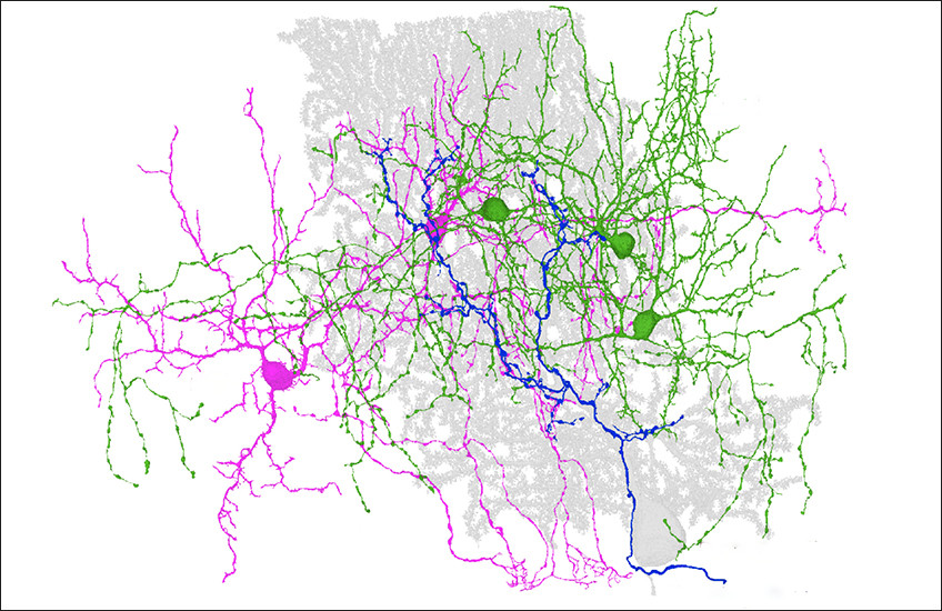
Homing in on one cell type split, the researchers focused on characterizing a cell category known as molecular layer interneurons (MLIs). Based on microscopy from the late 1800s, scientists had divided these cells into two types — basket and stellate cells — based on their appearance. However, RNA-Seq told a different story: It divided MLIs into two categories, which the researchers named MLI1s and MLI2s, based on starkly different gene expression. While MLI2s all looked like classic stellate cells, MLI1s looked like either basket or stellate cells.
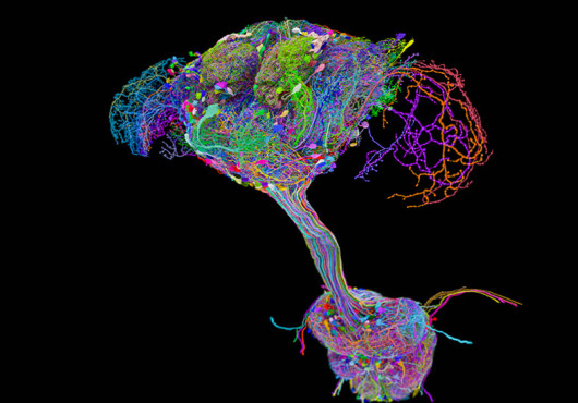
They then formed a powerful collaborative team to clarify the roles of MLI subtypes: Wei-Chung Allen Lee, associate professor of neurobiology at HMS and of neurology at Boston Children’s Hospital, used serial electron microscopy to provide insight into circuitry; Court Hull, associate professor of neurobiology at Duke University School of Medicine, recorded electrical activity from cells to determine how they function during behavior; and Nicolas Brunel, adjunct professor of neurobiology at Duke University School of Medicine, applied computational approaches to provide insight on cerebellar circuitry.
When the team investigated MLI function, they found another surprise. These cells were thought to inhibit Purkinje cell firing, creating a barrier for synapse formation. While that was true for MLI1s, MLI2s actually inhibited MLI1s, they published in Neuron in 2024. Thus, by indirectly encouraging Purkinje cell activity, MLI2s ultimately foster synapse plasticity in Purkinje cells, according to a 2026 study the team published in Nature.
Much more to explore
Regehr and colleagues continue to build on these and other findings about the cerebellar cortex, seeking to better understand the individual roles of each newly discovered cell type and how they function as a whole.
“We’ve known the architecture of the cerebellar cortex for well over 100 years and still don’t fundamentally understand it,” Regehr said. “I get up every morning thinking that we can make advances toward understanding what people thought was a relatively simple brain structure. Every time we look at it, it’s more interesting.”
Authorship, funding, disclosures
Macosko and Court A. Hull are co-authors of the review article. The authors acknowledge support from the Lefler Foundation and the Stanley Center for Psychiatric Research at the Broad Institute.