At a glance
New study traces how a protein essential for hearing evolved from an ancient ion channel into today’s exquisitely sensitive sound sensor.
Researchers pinpointed a small loop on the protein that appeared with the first vertebrate inner-ear hair cells, evolved further in mammals, and now is the site of many mutations that cause hereditary hearing loss.
Findings offer new clues to how hearing works at the molecular level and could help guide future therapies.
In the intricate machinery of the inner ear, hearing begins with a protein that moves a few billionths of a meter up to 100,000 times per second.
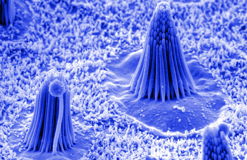
That protein, called TMC1, sits at the tips of sensory hair cells deep in the snail‑shaped cochlea. When sound waves move these microscopic hairs, TMC1 acts as a channel, opening and allowing charged particles to flow into the cell and trigger an electrical signal to the brain.
Without TMC1, that signal never starts. Mutations in the TMC1 gene are a well‑known cause of hereditary hearing loss in humans.
Because of this central role, TMC1 is an attractive target for researchers designing gene therapies aimed at restoring hearing. Several groups are testing ways to supply working copies of the gene or fix harmful mutations.
For these efforts to be safe and effective, scientists need to know in detail how TMC1 is built, how it opens, and which parts of the protein are most sensitive to change. However, the hair-cell system that includes TMC1 is so complex, sensitive, and hard to access that it is notoriously difficult to take apart and study directly.
A new study, published March 25 in Current Biology, begins to answer those questions by looking back in time.
In it, a team led by Harvard Medical School neurobiologists identified a tiny extracellular loop on the TMC1 protein as a crucial part of the channel’s gating mechanism, helping to control when it opens. They also found that the loop – which is known to be the site of many of the mutations to TMC1 that can cause deafness in humans – has been repeatedly refined by evolution over millions of years.
Tracing TMC1 through evolution
Senior author David Corey, the Bertarelli Professor of Translational Medical Science in the Blavatnik Institute at HMS, and colleagues combined three main approaches.
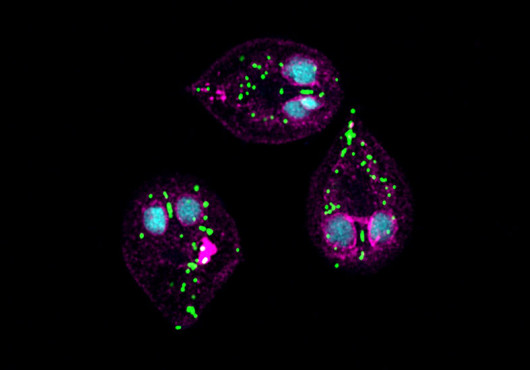
First, they used comparative genetics to search genomes from plants, fungi, and animals for TMC-related genes, align their sequences, and build an evolutionary family tree. This allowed the team to trace TMC1’s evolution from an ancient protein found in simple organisms into the finely tuned sensor that supports modern hearing. They saw when new branches of the TMC family appeared and which parts of the protein changed most as hearing became more specialized.
Second, they used artificial intelligence-based tools to predict how different versions of TMC1 fold into three dimensions. Comparing these structures showed that while TMC‑like proteins share a common core across species from single‑celled organisms to animals, one piece of TMC1 stands out in vertebrates, especially in mammals — a small loop that sits just outside the cell membrane and arches over the opening of the channel, where it contacts a partner protein called TMIE.

“What really jumped out was this small loop on the outside of the protein,” said Nurunisa Akyuz, former HMS research associate in neurobiology and co-first and co-corresponding author on the study. “This extracellular region evolves alongside vertebrate hair cells and continues to change in mammals, suggesting it helps tune the channel’s function.”
Initial structural and functional observations in the Corey lab pointed to this region, which the team then explored in depth through evolutionary analyses in collaboration with colleagues in the Department of Molecular Biology at Harvard University.
The analyses showed that this loop appeared when the first vertebrate hair cells evolved and that later, in mammals, that same region accumulated more changes than the rest of the protein. This pattern suggests that evolution kept tuning the loop to meet the growing demands on hearing, the authors said.
“All vertebrates use TMC1 in hair cells,” Corey said. “What is special about mammals is not that they invented a new protein, but that they refined an existing one, likely for high‑frequency, high‑sensitivity hearing.”
Third, the researchers tested these ideas directly in the lab. They engineered precise changes in the DNA that encodes the loop, delivered the altered TMC1 genes into the hair cells of a mouse model that normally lacks the TMC1 protein, and then measured the resulting electrical currents using sensitive electrodes. This allowed them to see how each mutation changed the way the channel responded to movement.
The tests showed a clear effect: Cells now needed larger movements of the hair bundle to open the channel. Adjusting this tiny external loop had changed TMC1’s sensitivity to sound.
“The loop is the part that appears when hair cells first evolve, keeps changing in mammals, and now turns out to influence how the channel opens,” Akyuz said.
From evolution to therapy
Taken together, the findings point to the loop as a key control element that helps set how easily TMC1 opens in response to tiny forces. They also identify it as a focal point where evolution, function, and human disease all intersect, offering a roadmap for interpreting deafness mutations and for designing future gene-based therapies.
“It is an extraordinarily sensitive protein,” Corey added. “Humans can hear very, very quiet sounds, and we think these evolutionary changes helped the channel open with even tinier changes in force.”
Authorship, funding, disclosures
Trey Scott, Corena Loeb, and Bifeng Pan are co-first authors with Akyuz. Additional authors include Yaqiao Li, Charles Phillips, and Nicholas Bellono.
This study was supported by the National Institutes of Health (grant R01DC000304), an American Hearing Research Foundation Discovery Grant, and a Harvard Brain Science Initiative Postdoctoral Pioneers Grant. The Boston Children’s Hospital Research Viral Core produced AAV vectors.