At a glance
An AI framework extends scientists’ ability to study how different parts of the brain communicate with and influence one another during behaviors such as memory formation.
The framework, called CURBD, has uncovered previously unknown brain-wide circuitry.
CURBD is available to labs worldwide and can be applied to existing datasets, allowing researchers to make new discoveries without designing new experiments.
The future of federally funded research at Harvard Medical School — supported by taxpayers and done in service to humanity — remains uncertain. Learn more.
For decades, neuroscientists have been able to record which parts of the brain show increased electrical activity during a behavior, such as when we form a memory or make a decision. But scientists haven’t been able to determine how those regions are communicating with and influencing one another, nor identify which regions serve as the main orchestrators.
A new artificial intelligence framework called Current-Based Decomposition, or CURBD, can now make that distinction. Developed by Harvard Medical School researchers and their colleagues, the tool is described August 7 in Neuron.
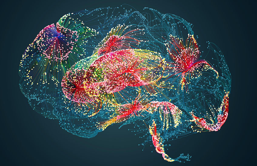
“No brain region works on its own. They’re all constantly sending signals to each other, shaping each other’s activity,” said Kanaka Rajan, associate professor of neurobiology in the Blavatnik Institute at HMS and the study’s co-senior and corresponding author. “CURBD lets us see those conversations like never before — which region is directing which, and how powerfully.”
The ability to map the brain’s internal communication has broad implications. For basic scientists, it opens a new window into how neural circuits give rise to thought and behavior. For clinicians, it raises the possibility of identifying, with much greater precision, which brain circuits go wrong in conditions like depression, memory loss, and movement disorders.
The paper represents more than six years of work and draws on collaborations with researchers at the Université de Montréal, Stanford University, the Icahn School of Medicine at Mount Sinai, and Cedars-Sinai Medical Center.
A new approach to brain mapping
Most existing tools, including newer AI-based approaches, tell scientists which brain regions are active and how active they are or what types of neurons are generating a given signal. They cannot reveal what is causing those neurons to behave that way nor how activity in one region is influencing activity across the rest of the brain.
CURBD addresses that gap by working backwards from the recorded activity of neurons to uncover what signals arriving from other regions drove them to fire in the first place.

To do that, Rajan, who is also a founding faculty member of the Kempner Institute for the Study of Natural and Artificial Intelligence at Harvard University, led a team to develop a step-by-step computational framework. A researcher feeds their neural recording data into CURBD, which uses it to train an AI model — a type of program called a recurrent neural network — until that model can accurately reproduce the patterns of electrical activity observed across the brain regions in the recording. Once the model has learned to mirror the brain’s behavior faithfully, CURBD analyzes its internal connections to read out which regions were sending signals to which others, how strong those signals were, and exactly when they arrived.
“If we’ve built something that walks like a duck and quacks like a duck, then the operating principles inside it are likely to match those in the real brain that we can’t otherwise access,” said Rajan — meaning that a model that perfectly reproduces a brain’s behavior may also reveal how that brain actually works.
New insights into depression and memory
To demonstrate the approach, the team applied CURBD to neural recordings collected across multiple species, including mice, nonhuman primates, and humans, and across a range of behaviors and disease-relevant conditions.
In several cases, CURBD extracted new insights from recordings that collaborating labs had originally gathered for entirely different purposes.
The findings shed new light on the brain circuits underlying conditions like depression and memory loss, with direct implications for how scientists understand and treat both.
In one set of experiments, the team applied CURBD to data from a well-established animal model of depression, in which subjects exposed to inescapable stress gradually stopped moving — a state analogous to the learned helplessness seen in human depression. Scientists had long suspected that a brain region called the habenula drives this behavioral shutdown by signaling to the raphe nucleus, which then releases serotonin.
CURBD confirmed part of that picture, but also revealed a second, unexpected mechanism. As subjects transitioned into passivity, a broad cortex-like structure called the telencephalon gradually increased its influence over the habenula in a distributed, brain-wide pattern. The circuit underlying depression-like states was far more complex than a simple relay.
“We thought we were going to find a light switch,” said Rajan of this experiment. “That’s not what happened.”
The finding has direct implications for understanding treatments like ketamine and deep brain stimulation, both used in patients with severe, treatment-resistant depression. Rajan and her team are now applying CURBD to compare these depression-related circuit patterns against recordings taken after ketamine is administered — work that could help explain why the drug succeeds at the level of brain circuits, even when years of other treatments have failed.
In a second set of experiments, the researchers applied CURBD to recordings from human participants — epilepsy patients with electrodes implanted across several brain regions at Cedars-Sinai Medical Center. They found that recalling a familiar memory is driven primarily by signals flowing from the frontal cortex to the hippocampus and amygdala — the brain’s memory centers — and that encountering a novel image recruits the entire network simultaneously.
This kind of insight into which regions are directing memory activity could prove valuable in research on conditions including Alzheimer’s disease.
Across experiments as different as stress responses in animals and memory retrieval in humans, CURBD consistently identified the specific regions driving each behavior — findings that would have been invisible to conventional tools. That consistency across nervous systems strengthened the team’s confidence that CURBD is revealing something fundamental about how brains are wired to communicate.
Looking ahead, with clinical potential
Because CURBD can be applied to neural recording data that already exists in labs around the world, researchers can use it to ask new questions of datasets they have already collected, without designing new experiments. In the case of the human data used in this study, the recordings had originally been gathered to help map epileptic seizures in patients undergoing evaluation for surgery. CURBD extracted new scientific insights from those recordings without any further involvement from the patients themselves.
The team has made the code for CURBD freely available, as part of the Kempner Institute’s commitment to open science.
Rajan sees the framework as a bridge between neuroscience and medicine — one that could help connect fields that have historically worked in silos. Both Parkinson’s disease and major depression involve a striking suppression of movement, yet neurologists and psychiatrists rarely share data or research frameworks, she said. CURBD, she believes, could provide a common analytical language to start those conversations.
“What I ultimately want is to write down something like a theorem for how brains work — a principle that holds across species, across diseases, across scales,” she said. “CURBD is a step toward that.”
Authorship, funding, disclosures
Karl Deisseroth of Stanford University is co-senior author, and Matthew Perich of the Université de Montréal and Quebec Artificial Intelligence Institute is first author of the study. Additional authors include Charlotte Arlt, Sofia Soares, Siyan Zhou, Manuel Beiran, Aaron Andalman, Tyler Benster, Megan Young, Clayton Mosher, Juri Minxha, Eugene Carter, Ueli Rutishauser, Peter Rudebeck, and Christopher Harvey.
This work was supported by the National Institutes of Health (grants R01MH110831, R01MH132064, National Institute of Mental Health BRAINS award R01MH110822 and R01MH118638, RF1DA056403, R01EB029858, NIH BRAIN Initiative R01EB028166, R01NS089521, and U01NS117839), the National Science Foundation (grants BCS-1554105, 1926800, and 2427124), the Gatsby Charitable Foundation (GAT3708), the James S. McDonnell Foundation (220020466), and the McKnight Endowment Fund for Neuroscience. Perich is supported by the Fonds de recherche du Québec - Santé (Artificial Intelligence in Health). Arlt is supported by a Mahoney Postdoctoral Fellowship. Soares is supported by a European Molecular Biology Organization Postdoctoral Fellowship. Rudebeck is the recipient of a young investigator grant from the Brain and Behavior Research Foundation. Harvey is the recipient of an NIH Director’s Pioneer Award (DP1MH125776).