
At a glance
Researchers have advanced colon-on-a-chip models to deepen studies of inflammatory bowel disease.
Chips included two types of colon cells taken from patients with IBD to mimic the intestinal wall. The team compared healthy and inflamed versions of those cell types from the same patients.
The models are the first to replicate certain disease features, such as increased likelihood of cancer development, and patient subgroups, such as pregnant women.
The work indicates that stromal cells are the primary driver of IBD and that pregnancy-related hormones directly affect disease severity.
Researchers have just made colon-on-a-chip devices even more useful for the study of inflammatory bowel disease (IBD), reproducing several aspects of the disease that have been missing in other lab models.
The work provides new insights into how IBD develops and progresses in specific patients, which could inform efforts to devise more effective and personalized treatments.
The study — published May 21 in Nature Biomedical Engineering — distinguishes which cell type is likely the primary driver of disease progression; demonstrates that hormones related to pregnancy and menstruation directly affect disease severity; and gives scientists the best opportunity yet outside of a patient or animal model to investigate why IBD cells are more likely than healthy gut cells to become cancerous.
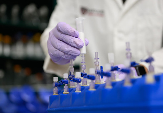
The international, multidisciplinary team was led by Donald Ingber, the Harvard Medical School Judah Folkman Professor of Vascular Biology in the Department of Surgery at Boston Children’s Hospital, founding director of the Wyss Institute for Biologically Inspired Engineering at Harvard University, and pioneer of organ chip technology. These microfluidic devices, lined with living human cells, mimic human organ systems. They provide more human-like alternatives to animal models and simple cell cultures for probing health and disease and conducting preclinical testing of potential new drugs.
Ingber said he hopes the team’s novel, personalized patient-to-chip approach will one day lead to more effective approaches to mitigate painful features of IBD and to prevent the formation of cancer in the intestinal tracts of people with IBD.
A new way to model IBD
IBD, which comprises the inflammatory conditions Crohn’s disease and ulcerative colitis, affects about 1.6 million Americans. Many people with IBD cannot be effectively treated, largely because scientists do not yet understand exactly what causes the increased inflammation, fibrosis (scarring), and compromised intestinal barrier that underlie this disease and its manifold symptoms, including severe abdominal pain, diarrhea, weight loss, rectal bleeding, anemia, anxiety, and depression.
Laboratory models of IBD could help untangle its causes and effects and advance new therapies, but such models are lacking, the authors said. Human in vitro models do not yet fully mimic the disease’s complex pathological features. Commonly used mouse models fall short of recapitulating human IBD because of differences in physiology, immunology, and sex-specific responses.

As a result, the exact processes in the intestinal system that trigger uncontrolled inflammation, loss of tissue integrity, and fibrosis “largely remain a black box,” explained Alican Özkan, first author of the study, who conducted the work as an HMS research fellow in the Ingber Lab and is now a senior scientist at AbbVie Immunology Discovery.
“Previously, researchers have used inflammatory molecules to induce these alterations without knowing precisely which cells actually produced them,” he said.
To provide insight into that question, clinicians at McGill University in Canada and Massachusetts General Hospital collected both healthy and inflamed colon tissue biopsies from patients with IBD.
The team grew and expanded epithelial (intestinal lining) cells from these samples in organoid cultures. They then cultured them in one of two parallel channels of a colon chip the size of a memory stick.
In the other channel, which is separated from the epithelial channel by a porous membrane, they created a stroma-like tissue (structural, connective cells) using fibroblasts derived from the same inflamed or unaffected patient tissue specimens.
The channels could communicate with each other by sending and receiving molecules that passed through the pores of the separating membrane.
This essentially created donor-specific IBD colon chips and healthy (control) colon chips that replicate the tissue-tissue interface seen in the actual intestinal wall.

Both the epithelial and the fibroblast tissue compartments were independently perfused with media, simulating the flow of intestinal fluids and blood, respectively.
Importantly, these human colon chips could also be subjected to peristalsis-like mechanical motions by cyclically stretching and contracting the membrane and adherent tissues in this flexible device, and human immune cells could be perfused or circulated through the device.
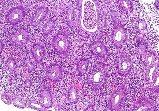
The IBD chips showed typical hallmarks of IBD, including a decreased height of the epithelial wall, leakage of unwanted molecules through the normally tight intestinal barrier, thinning of the secreted mucus layer that protects the intestinal wall from various insults, increased production of inflammatory molecules, and enhanced fibrosis (accumulation of fibrillar collagens by IBD stromal fibroblasts).
The gene expression patterns of cells in IBD chips also resembled those known for Crohn’s disease and ulcerative colitis.
Moreover, when the researchers perfused immune cells through the stromal channel, they migrated through stromal tissue, passed through the porous membrane, and entered into the epithelial tissue in IBD chips. This mimicked inflammation seen in vivo. Few immune cells did this in healthy chips.
Many of these IBD features were also exacerbated when the researchers applied peristalsis-like motions.
“Our human colon chips lined by living epithelial and stromal cells isolated from the same patients present an important breakthrough in IBD research because they enable us to control many different potentially contributing factors — including various cell types, hormonal exposures, and peristalsis motions, individually and in combination — which allowed us to gain new insight into key drivers of IBD development and progression,” said Ingber, who is also the Hansjörg Wyss Professor of Biologically Inspired Engineering at the Harvard John A. Paulson School of Engineering and Applied Sciences.
Insight into IBD drivers
The chip design enabled the team to address a long-standing question: Which intestinal cell type acts as the principal driver of IBD?
When the researchers paired inflamed epithelial cells with healthy fibroblasts on a chip, or healthy epithelial cells with inflamed fibroblasts, they found that the IBD-affected stromal fibroblasts were the major driver of many of the classic features of IBD, including barrier disruption and enhanced inflammation.
“Our study for the first time demonstrates that diseased fibroblasts are sufficient to cause healthy epithelial cells to take on many features of IBD,” said Özkan.
The approach also implicated mechanical forces associated with peristalsis motions.
Modeling pregnancy-specific IBD
The symptoms of IBD are often exacerbated in women, particularly during pregnancy, and the disease can raise risks of miscarriage, preterm birth, and low newborn birthweights. Physicians often warn women with IBD who seek to become pregnant to first ensure their disease is under control. But it hasn’t been clear whether pregnancy-related hormones exacerbate IBD in pregnant women directly by acting on the colon or indirectly by causing other changes in the body.

Ingber, Özkan, and colleagues used colon cells from female IBD patients in their chip model to investigate. The team flowed cocktails of hormones through the chips that mimicked either the menstrual cycle (estrogen and medroxyprogesterone acetate, or MPA) or the first trimester of pregnancy (estrogen, MPA, human chorionic gonadotropin, prolactin, and placental lactogen). Exposure to the hormone combinations enhanced inflammation and dramatically increased the level of fibrosis in the IBD chips, but not in healthy chips. This indicated that menstruation- and pregnancy-related hormones can have a direct impact on IBD severity.
“To my knowledge, this is the first model that has recapitulated in vitro the disease exacerbations that pregnant women with IBD often can experience,” said Ingber.
The findings offer a potential new avenue for finding therapeutic interventions for women with IBD who seek to become pregnant.
Studying colon cancer initiation in vitro
People with IBD have a higher risk of developing cancer in the intestinal tract than those without IBD. But how exactly this shift occurs is still enigmatic. An understanding of the changes in epithelial cells that drive the earliest stages of this transition would open new opportunities for researchers to develop cancer-preventing interventions. The team’s organ chips now make it possible to study how human cancers initiate within a relevant tissue and organ context in vitro.
To ask whether their model could be used to investigate the transition from IBD to cancer, the team exposed IBD chips and healthy chips to a carcinogen called ENU (N-ethyl-nitrosourea), working in collaboration with researchers at Queen Mary University of London and Cancer Research UK Cambridge Institute.
The IBD chips exhibited a significantly higher sensitivity to ENU than healthy chips. The levels of inflammatory molecules they produced were elevated, and signaling pathways known to be associated with cancer initiation became activated. The colon cells in the IBD chips also accumulated diverse types of gene mutations and chromosomal duplications, a result not seen in healthy chips.
“These findings are very exciting as, to our knowledge, they are the first to demonstrate early cancer progression in a preclinical human organ chip model,” said Ingber. “Our system enables studying the earliest stages of cancer formation within human tissues growing in an organ-relevant context in vitro.”
The findings once more implicated stromal fibroblasts and their aberrant interactions with epithelial cells.
The IBD-affected stromal cells, in particular, potently induced the expression of the early-stage colorectal cancer marker CEACAM5 in healthy epithelial cells exposed to the carcinogen, said Özkan. “This highlights the relevance of fibroblasts as important drivers of IBD-associated cancers as well as many other features of this disease.”
The work opens the door to more detailed investigations of the transition from healthy to cancerous tissue in the intestinal tract as well as the development of targeted interventions, particularly in IBD, Ingber said.
Adapted from a Wyss Institute news release.
Authorship, funding, disclosures
Additional authors on the study include Gwenn Merry, David Chou, Ryan Posey, Anna Stejskalova, Karina Calderon, Megan Sperry, Joshua Piatok, Viktor Horvath, Lorenzo Ferri, Emanuela Carlotti, Stuart McDonald, Douglas Winton, Rocco Riccardi, Liliana Bordeianou, Sean Hall, and Girija Goyal.
This work was funded in part by Cancer Research UK (award #C19767/A27145) and by the Wyss Institute as well as by National Institutes of Health training grants (5T32DK007199-44 and 5T32EB016652-10) and a Wyss Technology Development Fellowship. The authors thank the Harvard Digestive Diseases Center Organoid Core for providing L-WRN cell line-derived conditioned medium (P30DK034854).
Ingber holds equity in Emulate, chairs its scientific advisory board, and is a member of its board of directors. The other authors declare no competing interests.