Certain features in the spleen hidden within imaging scans may signal a person’s risk of coronary artery disease (CAD), according to a new study led by Harvard Medical School investigators at Massachusetts General Hospital.
The work helps answer the question of whether and how the spleen plays a role in coronary artery disease risk and suggests new targets for preventing and treating what remains the world’s leading cause of death.
The findings are published Sept. 9 in Science Translational Medicine.
Hidden in plain sight
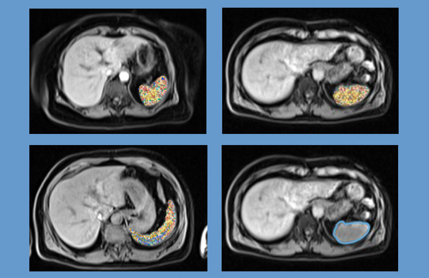
The research team looked to existing MRI scans of patients’ spleens to see what information could be extracted about this understudied organ’s association with plaque buildup in the arteries that supply the heart. Scientists have been trying to pin down the link for decades, given the spleen’s involvement in filtering the blood, protecting against infection, and regulating inflammation, blood pressure, and smooth muscle cells.
“Evidence increasingly ties the blood-forming system to heart disease, and the spleen is a central hub of that system, storing and filtering blood and producing immune and inflammatory cells,” said co-senior author Zhi Yu, HMS assistant professor of medicine at Mass General. “But the spleen is hard to assess through routine tests, so imaging is a powerful way to see nuanced changes in the organ and ask whether they reflect, or even forecast, disease.”
To investigate, Yu and colleagues leveraged artificial intelligence-based tools, abdominal imaging, and clinical outcomes data to identify splenic features relevant to coronary artery disease. They also performed genomic analyses to see if known CAD-associated genes are linked to splenic features seen in their imaging data.
The team’s research drew on information from 42,059 participants in the UK Biobank.

Ten of 107 splenic features observed on abdominal imaging scans were associated with CAD.
Genome-wide association analyses identified genes linked to both splenic features and CAD. These genes were often involved in processes such as inflammation, smooth muscle cell function, hypertension, and fat cell formation. Many of the associated variants fell in non-coding, regulatory regions of the genome. Two on chromosome 9 were associated with a nonuniform, irregular texture in the spleen and increased odds of CAD, independent of blood pressure, cholesterol, and other conventional risk factors.
Testing the findings in clinical care
To see how these general-population findings held up in patients, the team tested whether the UK Biobank patterns would also appear in people imaged as part of routine clinical care.
In 2,745 patients from the Mass General Brigham Biobank, most associations did not carry over. The authors suspect that this is in part because research cohorts like the UK Biobank tend to be healthier and scanned under uniform protocols, whereas clinical patients tend to have more complex medical histories and are imaged under widely varying protocols. The UK Biobank and Mass General Brigham settings may capture genuinely different biological states, they said.
“The cohort and protocol differences are likely a major driver of why our findings in the UK Biobank weren’t replicated in the clinical dataset,” said Yu. “But that doesn’t mean the findings are not generalizable. There is strong validation of the gene variants within the UK Biobank, and the genetics converge with independent observations in biology.”
Further work, such as applying more consistent imaging protocols to a clinical cohort, can strengthen confidence in the visual and genetic associations the team uncovered or weed out any that reflect chance, the authors said.
“These findings shed light on novel mechanisms linking the spleen to CAD, providing potential targets for therapeutic intervention to address this unexplored axis,” said first author Meghana Kamineni, HMS clinical fellow in medicine at Mass General.
Adapted from a Mass General Brigham news release.
Authorship, funding, disclosures
Pradeep Natarajan, HMS professor of medicine at Mass General, is co-senior author. Yu and Natarajan are also associate members at the Broad Institute of MIT and Harvard. Additional authors are Vineet Raghu, Zhanqing Hua, Haodong Tian, Buu Truong, Ahmed Alaa, Art Schuermans, Sam Friedman, Christopher Reeder, Romit Bhattacharya, Peter Libby, Patrick T. Ellinor, Mahnaz Maddah, Anthony Philippakis, and Whitney Hornsby.
This research was funded in part by the Harvard-MIT Health Sciences and Technology program, the HMS Office of Scholarly Engagement, the American Heart Association (Career Development Award 935176, 18SFRN34110082), a Harvard Catalyst K12 Award, Bayer AG, Fondation Leducq (14CVD01, TNE-18CVD04), the National Institutes of Health (1K01HL168231, 1R01HL092577, K24HL105780, 1R01HL134892, 1R01HL163099-01, R01AG063839, R01HL151627, R01HL157073, R01HL166538, 1K99HG012956-01, R01HL142711, R01HL127564, R01HL148050, R01HL151283, R01HL148565, R01HL135242, R01HL151152, and R01DK125782), the RRM Charitable Fund, the Simard Fund, and Massachusetts General Hospital through the Paul and Phyllis Fireman Endowed Chair in Vascular Medicine.
A full list of disclosures can be found in the paper.