The future of federally funded research at Harvard Medical School — supported by taxpayers and done in service to humanity — remains uncertain. Learn more.
Researchers at Harvard Medical School and the University of California San Diego have sped up a powerful imaging technique enough to map, for the first time, large volumes of brain tissue in nanoscale detail. The approach reveals fine structures that brain wiring maps, known as connectomes, can miss.
The work, described November 19 in Nature Methods and supported in part by federal funding, promises to help scientists build more complete diagrams of how cells in the brain are connected. This, in turn, could deepen understanding of nervous-system development and certain brain diseases.
“We can now see brain structures that we haven’t been able to see before in connectomics data,” said co-senior author Wei-Chung Allen Lee, associate professor of neurobiology at HMS and HMS associate professor of neurology at Boston Children’s Hospital.
“We hope this work will help us understand how nervous systems are built; what changes with age; and what goes awry in neurodevelopmental, cognitive, and neurodegenerative disorders,” Lee said.
Neuroscientists build connectomes to understand how neurons work together to produce thought and behavior.
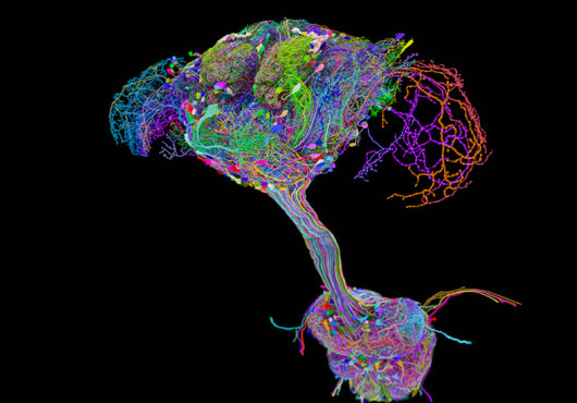
Some research efforts chart the brain’s major highways using MRI scans. Others zoom in, using electron microscopy to trace individual neurons and the synapses where they meet, as in a recently completed connectome of the fruit fly brain and nerve cord that Lee co-led with researchers from around the world.
However, even these fine-grained maps give up some detail when imaging large amounts of tissue. Electrical synapses, also called gap junctions, are among the key structures they can miss.
To capture that detail, the team turned to electron tomography, a technique that builds 3D images by viewing a sample from multiple angles. Electron tomography is usually slow because it requires taking hundreds of images of the same area.
The researchers found that they could use about 50 times fewer images than standard electron tomography, on tissue sections roughly 20 times thicker than usual, and still achieve 3D resolution of about 15 nanometers or finer.
Applying the technique to tissue from the mouse cerebellum, the team saw electrical synapses, which let signals pass almost instantly between neurons and are thought to help synchronize network activity.
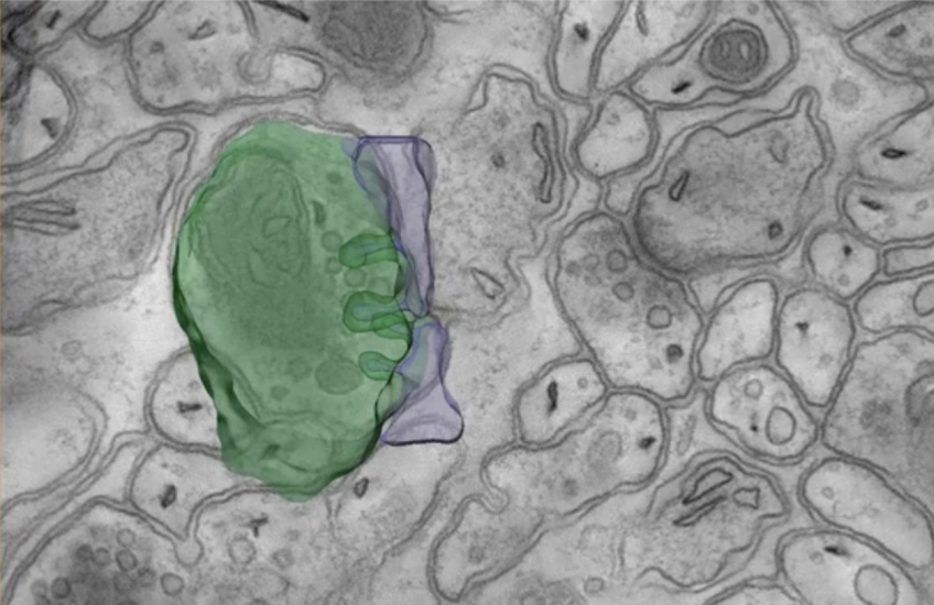
They also saw tiny protrusions extending between neurons and neighboring support cells called glia. The researchers say these structures appear to be involved in one cell taking up material from another.
“We’re seeing uncharacterized structures, suggesting that neurons are ‘nibbling’ their neighboring cells,” Lee said.
In addition, the team traced nerve fibers through a 3D block of tissue to the points where those fibers connect with other neurons.
Scaling the approach to an entire mammalian brain will take more work, including automating how tissue samples are handled, the researchers said. That is a long-term goal for the team.
Authorship, funding, disclosures
Aaron T. Kuan, a former postdoctoral fellow in the Lee Lab who is now at Yale School of Medicine, and Sébastien Phan are co-first authors of the study. Mark Ellisman is co-senior author. Additional authors include Kevin M. Delgado, Keun-Young Kim, Mason R. Mackey, Minsu Kim, Xiuye Chen, and Steven T. Peltier.
This work was supported by the National Institutes of Health (grants RF1MH129261, U01NS142013, and K99EB032217), with startup support to Kuan from the Yale School of Medicine Department of Neuroscience and Wu Tsai Institute.
Harvard University filed a patent application regarding GridTape (WO2017184621A1) on behalf of inventors, including Lee, and negotiated licensing agreements with interested partners. All other authors have no competing interests.