At a glance
An international collaboration co-led by Harvard Medical School researchers at Dana-Farber Cancer Institute has created 665 lab-grown models of patient tumors representing 27 cancer types and has made them publicly available.
The collection, annotated with clinical information and molecular datasets, roughly doubles the number of such models available to accelerate cancer research and development of new treatment strategies.
A team led by investigators at Harvard Medical School, Dana-Farber Cancer Institute, the Broad Institute of MIT and Harvard, and other collaborating institutions around the world has developed 665 next-generation cancer models — lab-grown cells derived from patients’ tumors — across 27 common and rare forms of cancer.
The resource, which the team has made publicly available, doubles the number of such models available for scientists to use to accelerate advances in cancer research and treatment.
Details of the work were published Aug. 5 in Nature.
The models are annotated with clinical information from patients, including outcomes and treatment history, as well as molecular data such as genomic sequencing information. It is the first time that such a large, annotated collection of patient-derived models has been made public all at once and validated as being reliable and faithful to the original samples.
“This is likely to be everyone on the team’s most important contribution to cancer biology in their career,” said co-senior author Keith Ligon, HMS associate professor of pathology at Dana-Farber and founding director of the institute’s Center for Patient Derived Models, which helped create many of the models in the collection. “The resulting resource enables new research at a large scale and for many years into the future.”
Addressing long-standing limitations
Researchers rely on lab-grown models of patient tumors because they offer a controlled way to study how cancers grow and respond to different treatments. But in the past, these models have not always remained biologically stable over time.
In the last decade, investigators at Dana-Farber and elsewhere have developed so-called next-generation methods to make these models, using cell cultures, plating, and growth methods customized for specific types of cancer. In these models, the biology of the tumor remains stable and responds to stimuli in ways that are representative of the original patient tumor. Researchers can reliably use them for years.
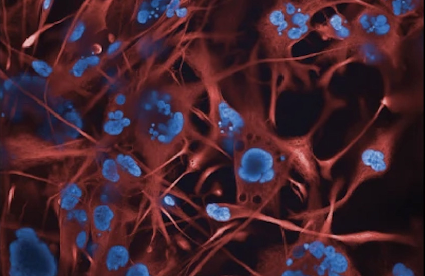
The models in the new collection include 3D organoids, which are miniature, lab-grown structures resembling tumors; 3D spheroids, which are tight clusters of cancer cells; and 2D cell lines, which are cancer cells grown as a flat layer in a dish.

They represent both adult and pediatric cancers, including colorectal, pancreatic, lung, and brain tumors, among others. More than 20 percent of the models represent rare cancer types, including types for which there were previously just one or two available models.
The collection includes 168 models from patients who had already received treatment, such as immunotherapy, targeted therapy, chemotherapy, or radiotherapy. It also includes 318 models of samples that were collected before patients began treatment.
An international effort
The models were created as part of the Human Cancer Models Initiative (HCMI), a large-scale international collaboration among the National Cancer Institute of the U.S. National Institutes of Health, Cancer Research UK, Wellcome Sanger Institute, and Hubrecht Organoid Technology. HCMI aims to create 1,000 next-generation patient-derived models that can be shared with the global community.
Approximately 2,800 patients receiving care at Dana-Farber and other locations in the United States, the United Kingdom, Italy, and the Netherlands consented to contribute tissue and data related to their cancer biology and treatment for this work. Samples were used to generate models that went through a rigorous validation process to ensure each was a faithful replica of the original.
Models that passed validation were then sequenced and analyzed to capture molecular data in a uniform way across the collection.
Researchers can access the models through the American Type Culture Collection, ordering them for lab experiments or obtaining their associated clinical and molecular data online.
Accelerating cancer research
Data from the models are already supporting cancer research efforts.
For example, HMS investigators at Dana-Farber used the models to expand the Cancer Dependency Map, a Broad Institute-based resource that uses CRISPR-based screening to identify new cancer vulnerabilities that therapeutics can target. Adding data from the new models expands the range of genetic and molecular subtypes represented in the screens and allows researchers to study models with gene expression and cell states that were not captured in earlier model systems.
“These new models and resources together represent a sea change advance in the tools available to the world, so we have what we need to fight cancer,” said Ligon. “They will be essential for generating the deep data needed for AI to help us unlock new treatments and break down barriers to rapidly help patients.”
Adapted from a Dana-Farber news release.
Authorship, funding, disclosures
Additional co-senior authors include Mathew J. Garnett of the Wellcome Sanger Institute, David A. Tuveson of the Cold Spring Harbor Laboratory Cancer Center, Andrea Califano of the Columbia University Irving Medical Center, Paul T. Spellman of the University of California Los Angeles, Daniela S. Gerhard of the Center for Cancer Genomics, Louis M. Staudt of the National Cancer Institute, and Jesse Boehm of the Broad Institute. Co-first authors are Dina ElHarouni, Mushriq Al-Jazrawe, Seongmin Choi, Merve Dede, Toshinori Hinoue, Sean A. Misek, Heeju Noh, and Luca Zanella. A full list of authors can be found in the paper.
Supporters of the work include the NIH/National Cancer Institute; Wellcome Trust; Cancer Research UK; Koch Institute Support; German Research Foundation; U.S. Department of Defense; Leidos Biomedical Research Inc; Lustgarten Foundation; Cold Spring Harbor Laboratory; Thomspon Foundation; Pershing Square Foundation; and Simons Foundation International. A full list of funding sources is available in the paper.
A full list of disclosures can be found in the paper.