At a glance
Study shows that two sensors used in brain research to detect different signaling molecules can report both signals.
The researchers advise caution interpreting data in certain circumstances and offer solutions to avoid mix-ups.
The findings could reshape how the sensors are applied in studies of Parkinson’s disease and other neurological conditions.
The future of federally funded research at Harvard Medical School — supported by taxpayers and done in service to humanity — remains uncertain. Learn more.
Cells in the brain communicate in different ways. The classical mode involves tight connections between individual cells that signal only to each other. But over the last decade, new tools have allowed scientists to focus increasingly on other, more diffuse systems, in which brain chemicals called neuromodulators communicate with many different cells over longer time frames.
Researchers at Harvard Medical School are now reporting that fluorescent sensors commonly used in this research can mix up two neuromodulators: dopamine and norepinephrine.
The discovery has important implications for studies of brain function, behavior, and disease. Abnormal signaling by dopamine and norepinephrine can drive symptoms seen in neurological disorders, including Parkinson’s disease, and obtaining accurate readings of each chemical at different times and locations in the brain is critical for understanding these conditions and developing interventions.
“For a mechanistic understanding of the physiology and pathology of these systems, it’s really important to know what these sensors are actually detecting,” said Pascal Kaeser, professor of neurobiology in the Blavatnik Institute at HMS and senior author of the paper, published Aug. 5 in Nature Neuroscience. “We found that, depending on which region of the brain you study, the norepinephrine sensor can report dopamine and the dopamine sensor can report norepinephrine.”

In addition to calling researchers’ attention to circumstances in which the sensors can produce false positives, the work offers insight into the complex underlying biology of neuromodulatory systems.
Different neuromodulators, similar signals
Neuromodulators shape how the brain responds to stimuli from within and outside the body, shaping behavior. Recently, scientists have developed tools to study neuromodulators in real time in live cells. Called G-protein-coupled receptor (GPCR)-based fluorescent sensors, they light up when particular neuromodulators bind to them on the surface of a cell.
GPCR-based sensors are fine-tuned to report one neuromodulator at a time. Kaeser and colleagues’ findings reveal that these sensors may fail to report the complexity of signaling in parts of the brain where multiple neuromodulators can act.
First author Ricardo C. López, a Harvard Kenneth C. Griffin Graduate School of Arts and Sciences PhD student in the Kaeser Lab, spearheaded the study. López was planning to measure norepinephrine release in the dorsal striatum, a part of the brain that coordinates habitual movements (such as typing or riding a bike), when he noticed something peculiar: The norepinephrine sensor signal strongly resembled that of dopamine.
The apparent mix-up was plausible given that dopamine and norepinephrine have nearly identical structures, though they do different things in the body. Norepinephrine, also known as noradrenaline and often referred to as the fight-or-flight chemical, prepares the body for action and helps keep the mind focused and alert. Dopamine, by contrast, helps the brain recognize rewarding experiences and identify which behaviors are worth repeating.
The possibility that the sensor was reporting two distinct neuromodulators “started ringing some alarm bells,” López said.
Scrutinizing brain signals
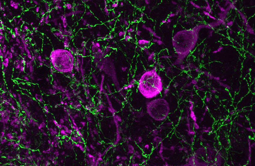
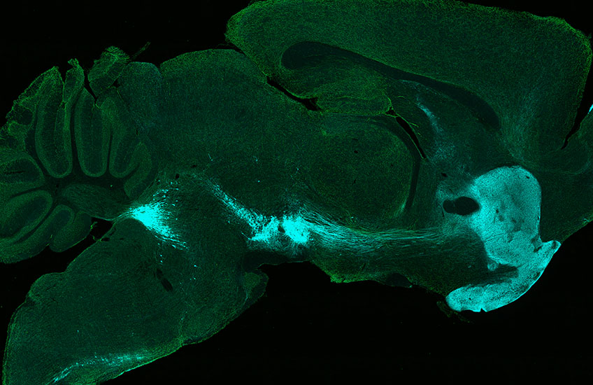
To investigate, López launched a series of experiments in mice designed to figure out what the sensors were reporting in different regions the brain. First, he used antibody staining to quantify and map the density of dopamine- and norepinephrine-producing nerve fibers in the dorsal striatum and in the motor cortex, a region that initiates voluntary movements.
According to his results, the dorsal striatum contained roughly 500 times more dopamine-producing fibers than norepinephrine-producing fibers, while the motor cortex showed an opposite pattern, with many norepinephrine fibers and very few dopamine fibers.
Next, López introduced genetic instructions into the animals’ brains to make either the norepinephrine or the dopamine GPCR-based sensor. Both sensors showed robust responses in the dorsal striatum and the motor cortex. That alone was a striking finding, given the innervation patterns observed in the mapping part of the study.
The decisive evidence came when López knocked out the cellular machinery that drives dopamine release. When dopamine release was blocked, the norepinephrine sensor signal in the dorsal striatum almost disappeared.
“The norepinephrine sensor had really been reporting dopamine,” Kaeser said.
A complementary experiment showed that dopamine sensors can similarly report norepinephrine.
Signaling molecules typically bind their receptors with lock-and-key specificity. But dopamine and norepinephrine are so similar, López said, that the receptor for one can be triggered by the other. And in regions like the dorsal striatum, where dopamine fibers vastly outnumber norepinephrine fibers, “even a small amount of crosstalk among the sensors can become dominant,” Kaeser added. “Researchers need to be aware of this limitation.”
A word of caution
Kaeser and López note that findings from studies that use these GPCR-based sensors to correlate neuromodulatory activity with behavior can be affected by these new findings. In future work, they said, sensor readouts must be interpreted cautiously, and researchers should use controls to remove one neuromodulator at a time to ensure the readout data are correct.
“You could imagine a paper linking Parkinson’s symptoms to a loss of norepinephrine in the striatum, when, in fact the norepinephrine signal is just cross‑reactivity of the sensor reporting dopamine,” López said.
Apart from this technical conclusion, the research reveals that neuromodulatory systems are more interrelated than some of the literature presents, Kaeser said. Dopamine under certain conditions can activate norepinephrine receptors, and norepinephrine can activate dopamine receptors, “and the blurring of these systems is revealed by the mixed sensor responses and very true at the disease level as well,” he said. “These systems interact with each other a lot.”
Authorship, funding, disclosures
López is studying in the Harvard Program in Neuroscience (PiN). Additional authors of the study are Natalie Noble, Özge D. Özçete, Leonardo Silenzi, Xintong Cai, Gillian E. Handy, Jonathan W. Andersen, Tommaso Patriarchi, and Yulong Li.
This work was supported by the National Institutes of Health (grants R01NS103484, R01DA056109, R01NS083898, and R01DA058777), an HMS Neurobiology Spark Grant, a Harvard Brain Initiative Bipolar Disorder Seed Grant, and an HMS Goldenson Research Fund Award. López is the recipient of a National Science Foundation graduate research fellowship (DGE2140743); Özçete was supported by a Human Frontier Science Program postdoctoral fellowship (LT0004/2022) and is the recipient of a Harvard Brain Initiative Postdoc Pioneer grant; and Andersen is the recipient of a Stuart H.Q. and Victoria Quan Fellowship. The authors also acknowledge the Neurobiology Imaging Facility and the Core for Imaging Technology & Education at HMS.
Li is listed as an inventor on a patent application (PCT/CN2018/107533) describing GPCR-based probes. Patriarchi is listed as an inventor on a patent application (PCT/US17/62993) describing GPCR-based probes.