The skin’s ability to replace the tissue it sloughs off is controlled by a range of genes, and now a study identifies the ringmaster of those genes as p63, a close relative of the well-known tumor suppressor p53. HMS cell biologists report in the May 4 Cell that p63 maintains a steady pool of regenerative epithelial stem cells and that p63-mutant mice lose all stratified epithelial tissues because they simply run out of these cells. The findings underscore the specificity of stem cell regulation in various tissues and add to the understanding of the mechanisms of regeneration.
Previously, HMS cell biology professor Frank McKeon found that without p63, mice are born without skin, a phenotype he described in the April 22, 1999, Nature. The p63 mutants showed a loss of stratified epithelial tissues, including skin, and lived for only a few hours after birth. “The phenotype is unprecedented,” McKeon said. “No single gene contributes as much to epithelial morphogenesis.”
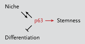
But the role of p63 in epithelial stem cells has been controversial, with some studies arguing that the protein contributes to differentiation of cells and others suggesting that it maintains a pool of regenerative cells. When the 1999 Nature paper was published, people had different explanations for the skinless phenotype. “If the gene isn’t operative, you don’t replenish basal cells,” said Christopher Crum, HMS professor of pathology at Brigham and Women’s Hospital and co-author on the Nature and Cell papers. “Others interpreted it as the inability to differentiate.”
Separated at BirthThe HMS researchers have gone on to show that p63 is at the crossroads of differentiation and “stemness,” as evidenced by the presence of markers of differentiation in the p63 mutants. “There was no question that the cells could differentiate,” Crum said.
So, why the conflicting views? It might be as simple as how skin tissue samples were collected. “In the p63-mutant mice, the epidermis forms during embryogenesis, but becomes highly fragile due to the loss of stem cells,” McKeon said. “This defective skin is lost during the process of birth.”
Filipa Pinto, a graduate student and co–first author on the Cell paper, did C-sections to obtain complete skin samples. “It seemed like the skin is peeling off; you can see the blood vessels beneath,” she said of embryonic tissue not obtained by C-section.
The researchers also studied the role of p63 in the thymus, a stratified epithelium that is shriveled in the p63 knockout. “The thymus is 10 to 20 times smaller than wild type at birth,” said Makoto Senoo, a research fellow and co–first author on the Cell paper. They wondered whether one of the two main cell types in the thymus—lymphocytes and epithelial cells—could be to blame.
Using a bone marrow transplant approach, Senoo showed that p63-mutant T cells could repopulate the thymus of mice lacking T cells. “The defect had to be in the epithelial cells rather than the lymphocytes,” he said.
Senoo then used the cell proliferation marker BrdU to show that wild type and mutant mice had the same number of proliferating cells in the thymus. “But somehow, the knockout gets smaller,” he said.
Having shown that low levels of epithelial cells seemed to be associated with a smaller thymus in p63-mutant mice despite the same amount of proliferating cells in mutants and wild types, the researchers tested whether the cells they were seeing were losing their ability to proliferate. They used epithelial stem cell cloning methods developed by Howard Green, the George Higginson professor of cell biology at HMS, to show that p63’s key function is to enhance the potential of stem cells to divide.
Seven days into cell culture, wild-type and p63-mutant cells were proliferating at the same rate. That was not so 12 days later, as non-proliferating cells became more and more abundant in the mutant tissue. The findings indicate an impairment in long-term proliferation in the p63 mutants, causing their cells to become terminally differentiated, or senescent, sooner. The mutants also had more markers of terminal differentiation, demonstrating a loss of proliferative potential.
End of the LineMcKeon and his research team used mathematical models to calculate the number of divisions the mutants might go through before becoming senescent. “We find that they can divide 15 times and then they’re at the end of their rope,” McKeon said. Meanwhile, wild types can divide 200 times. “The mutants are operating at low regenerative capacity.”
While the model is based on their findings from the thymus and the epidermis, the researchers suspect that the same low level of regeneration is happening in other stratified epithelial tissues, including the mammary and prostate glands, which also fail in the p63-mutant mice.
Interestingly, the gene only appears to be necessary for regeneration in stratified epithelial cells. “The epithelial stem cells of the single-layered GI tract do not express p63, and the p63 mutants do not have GI-tract problems,” McKeon said.
The specificity of p63 as a master regulator in stratified epithelia adds to an emerging literature of other tissue-specific master regulators of cell proliferation, such as the BMI-1 gene in hematopoietic stem cells and the PLZF gene in spermatogonia. “The fact that p63 is essential for these epithelial stem cells, while other master regulators have been identified for blood stem cells and spermatocyte stem cells, suggests a fundamental requirement for tissue specificity of these regulators that we don’t understand,” said McKeon. “Dissecting the genetic programs controlled by these regulators will tell us much about how stem cells function and how they go awry in cancer.”


