Felice Frankel likes to begin a talk to scientists by informing them she is not going to tell them anything they do not already know. Then the science photographer demonstrates to them, with telling use of color and composition, the rare power of a thoroughly considered image.
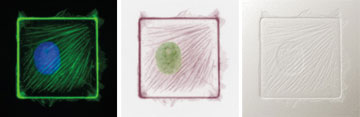
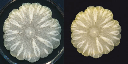
A dynamic and compelling image—be it of bacteria on a culture dish or a metaphor of genomic data printed as if lines in a telephone book—enhances more than its immediate subject, Frankel contends. An elegant picture helps scientists convey observations and processes to colleagues, converse with collaborators in other disciplines, capture the interest and support of the public and consider one’s own work in profoundly different ways.
“Everything I’m doing is all about how scientists can visually represent ideas and data,” said Frankel, a visiting scholar at the Wyss Institute for Biologically Inspired Engineering and an HMS research associate in systems biology. “This is nothing brilliant. It’s incredibly logical.”
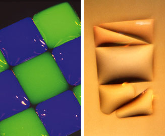
Some of the persuasive results are on display in her latest book, No Small Matter: Science on the Nanoscale, featuring the prose of coauthor and longtime collaborator George Whitesides, the Woodford L. and Ann A. Flowers University professor of chemistry at Harvard. Frankel created most of the images and also included contributions from HMS researchers such as Donald Ingber, Stephen Harrison, Roberto Kolter and their colleagues.
“Felice has changed the way scientists think about how to talk to one another and the public more broadly,” said Whitesides, who credits her in large part with the transformed role of journal covers from merely conveying information to peers to stimulating broader curiosity.
Visual ImpactLast year, Frankel relocated this visual exercise in communicating science from topics in chemistry, physics and engineering at Harvard and MIT to biology at HMS. In workshops at the Wyss Institute, Frankel will help researchers and postdocs of different disciplines transform their best images through a process of questions and collaborative responses. At the Medical School’s Systems Biology Department, she will push the process further into digital data collection and depiction.
“Scientists tend not to care what other people think,” said Ingber, founding director of the Wyss Institute and the Judah Folkman professor of vascular biology at HMS and Children’s Hospital Boston. “That’s fantastic. It’s why we can do things that are innovative and paradigm shifting. On the other hand, the only way our work can have an impact and change the world is to develop a wave of enthusiasm from others, and the only way to do that is to communicate, and the most powerful way to do that is visual.”
These conversations ultimately need to be an essential part of the scientific process. “One of the greatest concerns I have is that making a representation is, in itself, artificial,” said Frankel. “It’s a re-presentation. It’s not the thing. We have to be very careful when we show anything. There has to be some sort of transparent decision-making involved. By making a representation, we are, by definition, making a choice, an interpretation, which we hope, in the end, communicates. There is always part of it we don’t know. There’s always some sort of uncertainty, and we should inform the reader about that uncertainty.”
For more information, scientists and students may contact Felice Frankel at felice@felicefrankel.com.
Funding Sources: National Science Foundation, Harvard Division of Undergraduate Education


