Cell migration helps the body heal wounds. It allows the immune system to fight off nasty winter colds. And, more darkly, it allows tumors to form and spread. Because a cell’s ability to crawl about is such a fundamental part of cell biology, scientists have long been teasing apart the mechanisms that make motility possible.
New work from researchers at HMS and the Immune Disease Institute has identified an elegant bit of mechanics involving integrins, a family of cell surface proteins at the heart of a cell’s ability to connect to an outside surface and pull itself across. They found that simple mechanical forces tug an integrin open, springing it into an active conformation that allows the protein to grip an external surface as a tread on a tank does. The finding, described in the Dec. 26, 2008, Molecular Cell, may eventually provide insight into how to encourage cell migration when it is needed or stop it when it poses risk.
Integrin AnatomyThe work began with a feat of crystallography in the lab of Timothy Springer, the Latham family professor of pathology at HMS and a senior investigator at the IDI. Springer and Bing-Hao Luo, co–first author and a former research fellow in Springer’s lab, worked out a way to crystallize the full ectodomain of the integrin dubbed alphaIIb-beta3. Integrin proteins are large, forming an upside-down V with both feet dipping through the cell membrane and into the cytoplasm. The ectodomain is the part of the protein that is external to the cell, which, for this particular integrin, is the platelet.
A full integrin ectodomain had been previously crystallized, but several domains could not be seen because of disorder. The challenges in crystallizing the structure are manyfold; not only are integrins large, they have flexible components that need stabilization.
Luo managed to stabilize the protein and crystallize it, but challenges still remained. The structure contained clustered crystals. Co–first author Tsan Xiao broke the cluster into single crystals for analysis and built the first models of the protein in 2006. “It was not an easy structure to refine,” said Springer, who, along with co–first author Jianghai Zhu, spent another two years further refining it.
This work got Springer thinking about his next steps. His lab had learned much about the structure of integrins using other tools, such as electron microscopy. The evidence suggested that integrins change from a bent conformation with a closed headpiece to an extended conformation in a motion similar to the snap of a switchblade. Their new crystal structure reinforced this idea. However, the extended conformation can have either the closed, low-affinity or the open, high-affinity headpiece. What the researchers did not understand was what triggers the conversion of the headpiece from closed to open after the switchblade extends.
Scientists had been batting around two different hypotheses. In one, the integrin is thought to be “spring-loaded” and triggered to open by chemical signals, said Michael Dustin, a New York University pathologist whose work focuses on immune cell motility but was not an author on this paper. The other model posits that a force perpendicular to the cell surface pulls open the protein.
Forces at PlayWith the full ectodomain of the structure at his disposal, Springer had enough information to test the mechanical hypothesis. To do so, he used a technique called steered molecular dynamics, which uses computers to simulate the exertion of forces on a molecule. Zhu and co-author Chengzhong Zhang, both research fellows in pathology, used the technique to pull the head domain of the protein away from the plane of the cell surface. They expected this force to activate the integrin.
“It didn’t happen that way,” said Springer. The protein extended, but the headpiece stayed in its closed, low-affinity conformation. It was then that Springer realized he had forgotten an important piece of the puzzle.
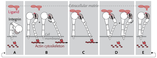
In addition to being pulled in a direction orthogonal to the cell surface, such as when two cells move apart from one another, proteins can also be pulled laterally. This happens, for example, during cell migration. In the cytoplasm, the filament network of the actin cytoskeleton forms at the leading edge of the cell. This network grabs onto the front foot of the integrin and drags the protein along the surface of the cell. The integrin then acts like a tread that grabs onto the extracellular matrix, enabling the cell to propel itself across it.
This lateral pull has long been known, said Springer. “But when people were thinking about integrin activation, they just hadn’t put the two things together.”
When Springer and his team used their molecular dynamics simulation to simultaneously tug orthogonally at the head of the protein and laterally on the leading foot, where the actin cytoskeleton holds on, the protein both extended and snapped open (see diagram). This conformational change alters the chemistry, creating a high-affinity binding site that extends up to 300 angstroms from the cell surface, farther than typical receptor binding sites.
“I had thought that activation would be very complicated and would involve a lot of intracellular pathways and regulators,” said Springer. “Certainly there are lots of chemical processes going on to activate the cytoskeleton and so on, but at the level of the molecule, it’s really a very simple mechanism for connecting ligand binding outside with binding to the cytoskeleton inside.”
This is an “exciting conclusion,” said Dustin. “It is a key insight that requires a whole view of the way an integrin works in a cell plus a view of the integrin at the atomic level at the same time.”
It is not clear yet how these simulations might play out in an actual cell. The work does, however, suggest that the “integrating function”—the heavy-sounding words often used to describe the way integrins combine internal signals with external ones—may be a simple mechanical switch. “When the cytoskeleton is attached, the integrins are adhesive. When it’s not attached, they’re not adhesive,” said Springer.
Springer and his team plan to demonstrate this mechanism at the cellular level. They also want to understand whether this mechanochemical machinery is a general principle that applies to all integrins.


