When the immune system detects a threat, cells communicate with one another through signals that activate inflammation. Scientists have long understood that these signals are relayed through receptors: proteins on or inside cells that detect danger and pass along instructions.
Now, Harvard Medical School researchers at Boston Children’s Hospital and Dana-Farber Cancer Institute have reported that inflammatory signals can continue without receptors, revealing alternative pathways for cell-to-cell communication and a potential new target for treating autoimmune disease.
The results published Sept. 23 in Nature.
“The discovery of this receptor-free signaling transduction underscores the value of exploratory science as a means to expand the lexicon of life,” said senior author Jonathan Kagan, the HMS Marian R. Neutra, PhD Professor of Pediatrics at Boston Children’s.
For decades, receptor-mediated signal transduction has been considered the model for how cells communicate inflammatory signals. In this process, receptors detect changes in the environment — such as other cells, infections, or cancers — and, once detected, bind to proteins within the cell’s cytoplasm that signal to induce changes in cell function.

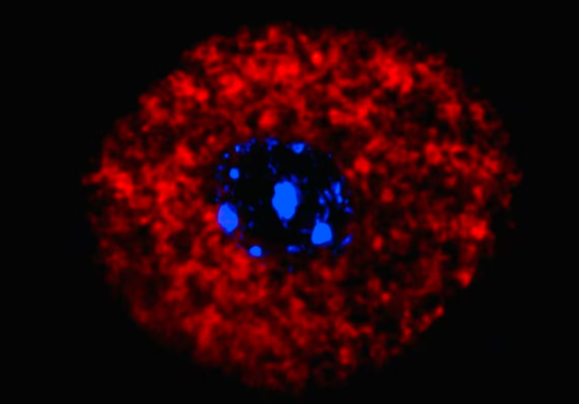
Using a combination of genetic, imaging, transcriptomic, and biochemical approaches with immune cells in a dish, Kagan and his team observed a different process.
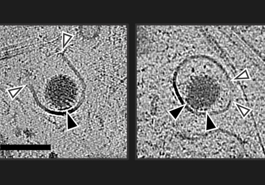
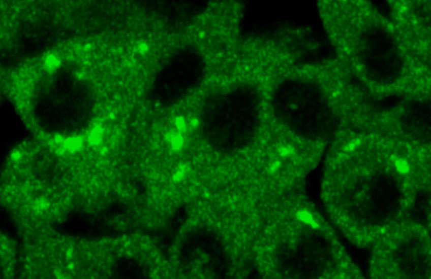
During inflammation, toll-like receptors (TLRs), proteins that help immune cells detect signs of infection, assembled signaling proteins but then released them. Contrary to dogma, the release did not stop the inflammatory process.
Rather, the receptor-free proteins were needed to assemble inflammation-stimulating organelles called myddosomes. Without the proteins’ release from the receptors, myddosomes could not form nor induce inflammatory gene expression.
The findings suggest that separating from the receptor is not the end of the signal, but a step that allows inflammatory signaling to continue.
The ability for cells to instigate functional changes without receptors acting as the middleman has both scientific and clinical implications. Since the Nobel Prize-winning discovery of TLRs as the driver of inflammation and immunity in the 1990s, scientists have worked to develop therapies that suppress TLR activity by interfering with receptors, with little success.
The discovery of receptor-free signal transduction may help explain that limited success. Because the myddosome can continue driving inflammatory responses after separating from the receptor, it may be a more effective target for therapies.
Indeed, in a mouse model, the researchers found that using a drug to disrupt myddosomes reduced levels of inflammatory proteins in the blood.
“New strategies to manipulate the TLR pathway, central to so many aspects of inflammation, are now possible,” said Kagan. “Our hope is to develop new classes of small molecules that manipulate myddosomes as a means of therapeutic treatment for autoimmune diseases, and beyond.”
Adapted from a Boston Children’s news release.
Authorship, funding, disclosures
Daniel Fisch, HMS research fellow in pediatrics at Boston Children’s in the Kagan Lab, is first author of the paper. Additional authors include Vaani Ohri, Eleni Anastasakou, Lucia J. Wesemann, Joon H. Choi, and Katherine E. Lindblad.
This study was supported by the National Institutes of Health (grants AI167993, AI116550, AI184490, AI198293, DK34854, and DK007260), a Human Frontier Science Program postdoctoral fellowship (LT0006/2022-L), an EMBO postdoctoral fellowship (ALTF 491-2022), a HRIA Charles A. King Trust postdoctoral research fellowship, and a Cancer Research Institute Irvington Postdoctoral Fellowship (CRI14367). The Nikon TIRF microscope used in the study was purchased with the support of the NIH (S10 RR027344-01).
Kagan receives compensation from and holds equity in Corner Therapeutics, Larkspur Biosciences, MindImmune Therapeutics, and Neumora Therapeutics. He reports that none of these relationships impacted this study. The other authors declare no competing interests.