 Because most human tissues do not regenerate spontaneously, advances in tissue repair and organ regeneration could benefit many patients with a wide variety of medical conditions.
Now a research team led by Harvard Medical School investigators at Beth Israel Deaconess Medical Center and Dana-Farber/Boston Children’s Cancer and Blood Disorders Center has identified an entirely new approach to enhance normal tissue growth, a finding that could have widespread therapeutic applications.
Their findings were published online July 29 in the Proceedings of the National Academy of Sciences.
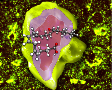
Tissue regeneration is a process that is not fully understood, but previous research has demonstrated that endothelial cells lining the insides of small blood vessels play a key role in tissue growth. It is also known that these endothelial cells generate chemical messengers called epoxyeicosatrienoic acids (EETs), which stimulate blood vessel formation in response to tissue injury.
In this new research, first author Dipak Panigrahy, an instructor in pathology at HMS and an investigator in the Beth Israel Deaconess Center for Vascular Biology Research, and his colleagues wanted to find out how EETs might participate in organ and tissue regeneration. To answer this question, they created seven different mouse models. The models focused on liver, kidney and lung regeneration; wound healing; corneal vascularization and retinal vascularization.
“We used genetic and pharmacologic tools to manipulate EET levels in the animals to show that EETs play a critical role in accelerating tissue growth, providing the first in vivo demonstration that pharmacological modulation of EETs can affect organ regeneration,” explained Panigrahy. Administering synthetic EETs spurred tissue growth in the research models; conversely, lowering EET levels—by either manipulating genes or administering drugs—delayed tissue regeneration.
The team also demonstrated that proteins called soluble epoxide hydrolase (sEH) inhibitors, known to elevate EET levels, promoted liver and lung regeneration. sEH is the main metabolizing enzyme of EETs.
“Our results offer a mechanistic rationale for evaluating sEH inhibitors as novel therapeutics for a number of human diseases such as hepatic insufficiency after liver damage and diseases characterized by immature lung development, such as bronchopulmonary dysplasia,” said Panigrahy, adding that the use of topical sEH inhibitors on the skin might also be useful for the acceleration of wound healing.
The researchers suspected that EETs were stimulating tissue regeneration by way of blood vessel formation, specifically by producing vascular endothelial growth factor (VEGF) to promote vessel growth. As predicted, when the investigators depleted VEGF in the mice, effects of EET on organ regeneration disappeared.
“Discovering EETs’ role could be of critical importance to help control the repair of liver, lungs and kidneys,” said senior author Mark Kieran, an HMS associate professor of pediatrics at Dana-Farber/Boston Children’s Cancer and Blood Disorders Center. “Since diseases of these organs are a major cause of morbidity and mortality in the North American population, the opportunity to modulate the regeneration of healthy tissue could have significant therapeutic implications for many patients.” These findings may also apply to conditions or physical defects that lead to the loss of specialized cells in other organ systems, such as the nervous system and the immune system.
The investigators stress that it will be important to determine whether EETs affect other factors besides VEGF in influencing tissue repair. Additionally, they add, the beneficial effects of EETs will have to be carefully weighed against their finding that direct administration of EETs can stimulate cancer growth in animal models. Several clinical trials that are currently testing the potential of sEH inhibitors for purposes other than organ regeneration or wound repair could offer valuable insights into the safety of elevating EET levels in patients.
“Although our work suggests synthetic EETs would promote wound healing after surgery, more clinical trials are needed to assess the potential benefits and possible risks of these novel lipids,” added co-corresponding author Darryl Zeldin, scientific director for the National Institute of Environmental Health Sciences, part of the National Institutes of Health.
This work was supported by grants from the National Cancer Institute (RO1CA148633-01A4); the Stop and Shop Pediatric Brain Tumor Fund; the C. J. Buckley Pediatric Brain Tumor Fund; the Children’s Hospital Boston Surgical Foundation and the Vascular Biology Program; the Robert A. Welch Foundation (GL625910); the Intramural Research Program of the NIH, National Institute of Environmental Health Sciences (Z01 025034 and Z01 050167); the National Institutes of Health (R01 GM088199; GM31278; R01 ES002710; R01 ES013933, and CA045548); and the NIEHS Superfund Basic Research Program NIH Grant P42 ES004699. The work was also supported through the Joshua Ryan Rappaport Fellowship and Howard Hughes Medical Institute Research Fellowship.
Adapted from a Beth Israel Deaconess news release.