Video: Kirchhausen lab
This article is part of Harvard Medical School’s continuing coverage of COVID-19.
In a first, scientists have captured on video all the steps a virus follows as it enters and infects a living cell in real time and in three dimensions.
They achieved the feat by using advanced imaging called lattice light sheet microscopy as well as chemical and genetic manipulation.
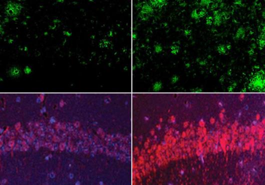
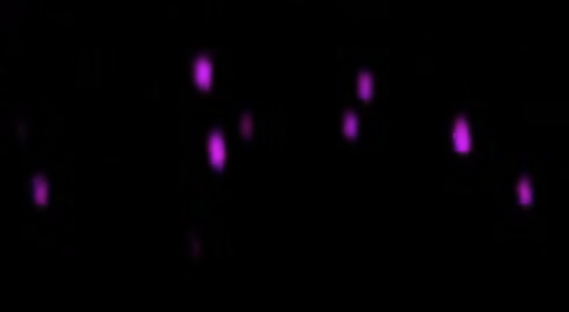
The first part of the video shown here follows a virus engineered to sprout SARS-CoV-2 spike proteins (labeled pink) as it is captured at a cell surface and engulfed by a cellular compartment called an endosome. The virus then fuses with the endosome membrane and injects its genetic material (labeled blue) inside the cell — the steps necessary to kick off a cycle of viral infection and replication.
The second part of the video shows many such viruses inside the cell. The video spans about 4 minutes, with snapshots taken every 4 seconds.
Enlightening, understandable, provocative
Solid reasons to read Harvard Medicine magazine