At a glance
HMS researchers have developed NoR-AI, a tissue-imaging pipeline that combines a custom microscopy method with AI-powered image analysis to capture fine details that existing histopathology methods miss.
NoR-AI provides unprecedented levels of detail and the ability to observe lipids, which are typically removed during tissue preparation.
The work could help researchers uncover new features associated with aging and disease and provide a basis for faster, more detailed clinical evaluation of tissues.
The future of federally funded research at Harvard Medical School — supported by taxpayers and done in service to humanity — remains uncertain. Learn more.
For centuries, researchers and clinicians have used microscopes to study the causes and effects of disease on tissues and help make diagnoses — the practice of histopathology. While the microscopes have certainly improved, scientists still rely on tissue preparation approaches that distort subtler features and entirely remove lipids and water-soluble materials. The result is an incomplete and imperfect view that cannot capture lipid data and other details that may be relevant to disease.
Researchers in the Blavatnik Institute at Harvard Medical School have developed a new imaging pipeline that overcomes these limitations and provides an unprecedented look into tissues.
The work could help researchers uncover new features associated with aging and disease and provide a basis for faster, more detailed clinical evaluation of tissues.
The pipeline, named NoR-AI, combines a custom microscopy method with AI-powered image analysis to capture high-quality, quantitative images that include lipids alongside proteins and water content. The technique requires little to no pre-processing of tissues and can be completed in 30 minutes, as compared to several days for traditional biopsy analysis.
In a federally funded study published Nov. 5 in Proceedings of the National Academy of Sciences, the researchers used NoR-AI to identify previously unrecognized fine-scale changes in a mouse model of acute kidney injury.
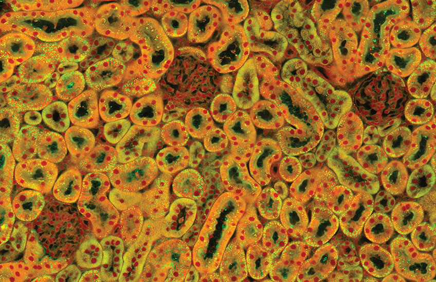
“We’re able to see into cells and tissue in ways that nobody could before,” said senior author Marc Kirschner, the John Franklin Enders University Professor of Systems Biology at HMS. “We can tell how much protein, how much lipid, how much water there is in these tiny little volume elements. And if you look at the larger picture, we can see unbelievably beautiful structures and cells all the way through the tissue.”
A new look at kidney damage
NoR-AI builds on an imaging process called normalized Raman imaging, or NoRI, developed by co-first author Seungeun Oh while she was a postdoctoral researcher in the Kirschner Lab. (Oh now runs her own lab at the University of California, San Diego.)
NoRI creates images using stimulated Raman scattering, a technique that measures how light changes as it interacts with different tissues. NoRI then computationally corrects for light-scattering effects that can interfere with the measurements pathologists and other researchers want to make.
In NoR-AI, the clear, quantitative images provided by NoRI are then analyzed by a convolutional neural network — a type of AI-powered image analysis. Because the NoRI images are so detailed, the neural network can pick out fine-scale distinctions that other methods miss.
“The problem with applying these neural networks to traditional histology images is that they have to see through so much noise to resolve features,” said co-first author William Trim, research fellow in systems biology in the Kirschner Lab at HMS. “With NoRI there’s no noise. It’s just the clean tissue, as it was in the body.”
With as few as two or three NoRI images of mouse kidneys, the neural network was able to identify three distinct tubule types; classify anatomical regions; and determine the sex of the mouse based on lipid and protein distribution in the tubules.
In collaboration with Joseph Bonventre, the HMS Samuel A. Levine Distinguished Professor of Medicine at Brigham and Women’s Hospital, the team also tested NoR-AI with a mouse model of acute kidney injury, capturing changes in lipid and protein organization over 25 days post-injury. They were able to characterize and quantify several known features of acute kidney disease and uncover new aspects of kidney damage and repair that had not been observed before, including how lipids are lost and gained in different areas over time.
“We have information in these NoRI images that hasn’t previously existed,” Trim said. “It can only be seen using NoRI combined with a neural network.”
“You wouldn’t have the lipid data without NoRI, and you wouldn’t spot these tiny changes without the neural network,” he said.
Uncovering the role of lipids
Although the team used the kidney as a proof of principle, NoR-AI has the potential to uncover new features and improve diagnostic precision in other tissues as well. Kirschner, Trim, Oh, and their colleagues are already using NoR-AI to investigate preclinical models of colon, liver, and prostate cancers and plan to expand into diabetic kidney disease and other areas.
NoR-AI can also tell the difference between different types of lipids, such as cholesterol, saturated fats, and unsaturated fats, making the tool potentially useful for deepening understanding of these lipids’ roles in inflammation and immune system response.
“Lipid is the terra incognita of histology,” Kirschner said. “But in the cases we’re looking at, we’re starting to see that the lipid amounts and distribution change significantly. That’s got us and a lot of other people excited.”
Authorship, funding, disclosures
Additional authors of this work include Mariia Diakova, Kseniia Petrova, Takaharu Ichimura, Ayumi Takakura, Ranit Karmakar, Simon F. Nørrelykke, and Leonid Peshkin.
This work was supported by the National Institutes of Health (grants R01AG073341, R01DK039773, and R01DK072381 and Maximizing Investigators’ Research Award R35GM145248).
Kirschner and Oh hold a patent for Normalized Raman Imaging (NoRI), US11971357B2.