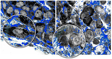
Cancer’s deadly journey into other parts of the body starts when normal processes that restrain growth go awry, allowing tumor cells to escape their primary site, invade tissue and multiply. New research suggests that when proteins that orchestrate cell architecture are absent, tumor invasion can be more pronounced and prognosis poorer for breast cancer patients.
These findings, published Nov. 13 in Cancer Cell, are based on experiments in cell culture and in mice engineered to develop human breast cancer, as well as an analysis of samples from breast cancer patients. While their discovery does not have immediate clinical relevance for cancer patients and their doctors, the finding inspires new avenues for exploring how cancer spreads.
“This work contributes to understanding one of the initial steps of cancer metastasis: invasion into the tumor microenvironment,” said Joan Brugge, Louise Foote Pfeiffer Professor of Cell Biology and head of the Department of Cell Biology at HMS.
Normally, when breast tissue expands during pregnancy to prepare for lactation, there is a dramatic branching of individual ducts to create a flowering of ductal systems that deliver milk to the baby. That highly controlled branching is a normal process that goes wrong in tumor development.
To find out how this process is corrupted, the scientists focused on profilin-1 and profilin-2, two proteins that are almost identical except for small differences in their amino acids and binding partners, which are, in turn, responsible for the varied behaviors of proteins. These two proteins play distinctive roles in building the cell’s skeleton, a structure that regulates multiple aspects of cell behavior, including cell migration, common in both normal and cancerous growth. Profilin-1 promotes dynamic membrane protrusions filled with branched networks of actin filaments, the chains of actin polymers that enable cells to move through tissue. In contrast, profilin-2 promotes the formation of distinct actin structures, made of actin bundles, which create a physical barrier that inhibits membrane protrusions.
Profilin-1 is widespread and well studied throughout the body, but profilin-2 has captured little attention in cancer biology because it appears most often in the nervous system. Taking a closer look at profilin-2, first author and postdoctoral fellow Ghassan Mouneimne found that to do its job of suppressing protrusions, profilin-2 depends on another protein, called EVL. This protein, a less-characterized ally of profilin-2, bundles filaments on the cell membrane in a way that prevents protrusion outside the cell and invasion into a tumor’s neighborhood.
Looking at samples from breast cancer patients and at publicly available data, the scientists observed that metastatic cancers show low levels of EVL, fitting their hypothesis that inhibiting EVL’s actin bundling promotes tumor spread. Published studies that followed cancer patients over time found that those whose tumors had lower EVL had worse outcomes.
“Profilin-2 and EVL collaborate to suppress the invasive behavior of cancer cells,” said Mouneimne. “Based on the statistical analysis we did, EVL may be a biomarker for metastatic potential in breast cancer.”
Before considering EVL as a biomarker or developing strategies for therapy, the scientists must determine what causes profilin-2 and EVL to fail. A genetic mutation could be the culprit, or an epigenetic alteration—a change in gene expression or some other modification—could be the trigger for their loss of function.
Profilin-2 and EVL are part of a complex story.
“It’s not just one pathway that drives invasive behavior in tumor cells,” Brugge said. “Any of a combination of different types of alterations will promote invasion. This work tells us about an important one.”
This research was funded by the Breast Cancer Research Foundation, the NIGMS Cell Migration Consortium, a gift from the Lee Jeans Foundation through the Entertainment Industry Foundation, UCSF/UC Berkeley Nanomedicine Development Center, the National Science Foundation, and NIH grants P01 HL059561, ROI GM61010, and NIH GM58801.


